
- Physical Examination
- Surgical Examination
- Ophthalmology
- Clinical Skills
- Orthopedics
- Surgery Videos
- Laparoscopy
- Pediatrics
- Funny Videos
- Cardiothoracic Surgery
- Nursing Videos
- Plastic Surgery
- Otorhinolaryngology
- Histology and Histopathology
- Neurosurgery
- Dermatology
- Pediatric Surgery
- Urology
- Dentistry
- Oncology and Cancers
- Anatomy Videos
- Health and Fitness
- Radiology
- Anaesthesia
- Physical Therapy
- Pharmacology
- Interventional Radiology
- Cardiology
- Endocrinology
- Gynecology
- Emergency Medicine
- Psychiatry and Psychology
- Childbirth Videos
- General Medical Videos
- Nephrology
- Physiology
- Diet and Food Health
- Diabetes Mellitus
- Neurology
- Women Health
- Osteoporosis
- Gastroenterology
- Pulmonology
- Hematology
- Rheumatology
- Toxicology
- Nuclear Medicine
- Infectious Diseases
- Vascular Disease
- Reproductive Health
- Burns and Wound Healing
- Other
Top videos
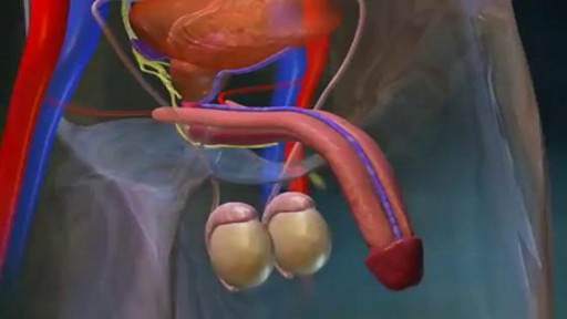

What Happens During an Erection?
In order to attain an erection, messages from the brain and other sense organs trigger the arteries of the penis to dilate. This allows an increased amount of blood to flow into three columns of spongy tissue in the penis.
As the arteries supplying blood to the corpus spongiosum and to the two larger columns, the corpus cavernosa, become filled with blood; the penis grows and becomes rigid. Pressure of the engorged tissue against the veins in the penis effectively traps blood within the penis until climax is reached or the sensation wanes.
What Are Penile Implants?
Impotence, or the inability to attain or maintain an erection, can be caused by a disruption at any stage in this process. Several types of penile implants are available that create an artificial erection. Two common types of implants are the semi-rigid malleable rod and the inflatable implant.
•The semirigid malleable rod is usually made of plastic with a core of flexible wire. These rods can be bent down to conceal the penis under clothing or raised to form an artificial erection.
•The inflatable implant is more complex and involves several working parts: a reservoir of fluid that is implanted into the abdomen, a pump system located in the scrotal sac near the testes, and two inflatable cylinders.
How Penile Implants Help Erectile Fuctioning
In order to attain an erection, the scrotal pump must be squeezed repeatedly to propel fluid into the penile cylinders. When an erection is no longer desired, a release valve is pressed on the side of the pump and the cylinders deflate.
Before Having Penile Implant Surgery
Persons considering these types of implants should speak with their physician or healthcare professional about possible risks and complications.
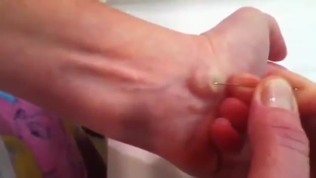
A ganglion cyst is a tumor or swelling on top of a joint or the covering of a tendon (tissue that connects muscle to bone). It looks like a sac of liquid (cyst). Inside the cyst is a thick, sticky, clear, colorless, jellylike material. Depending on the size, cysts may feel firm or spongy.
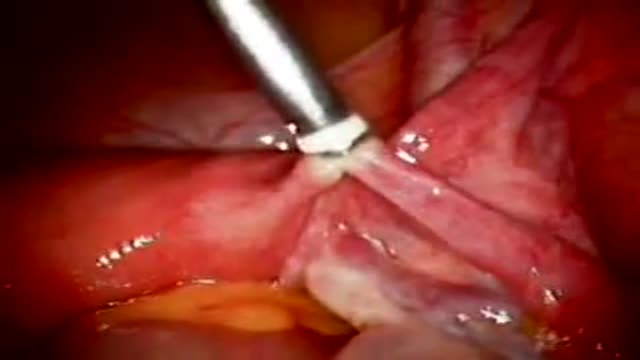
Tubal ligation using Fallope Ring

Professional Breast Exam
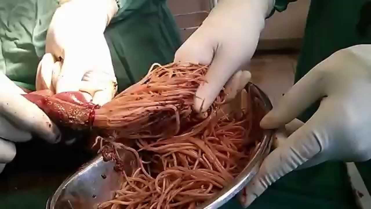
Watch that 3 Years Old Intestinal Worms Removal Surgery
Part 1: from Loyola Medical School, Chicago showing clinical examination of the neurological system.
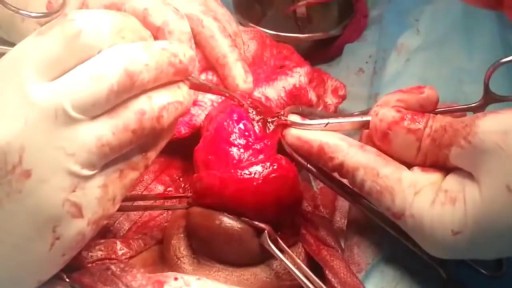
Watch that video of Broken Male Genital Repairing Surgery
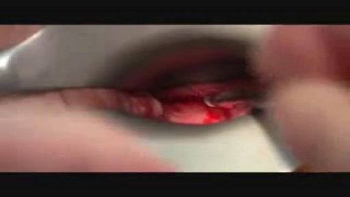
When both mucosa and stroma are parts of the suspect lesion, a deep biopsy is needed. The Cervicore is designed to harvest samples from the cervix and vagina with minimal collateral injury to the surrounding tissues. The procedure is easy with minimal discomfort to the patient.
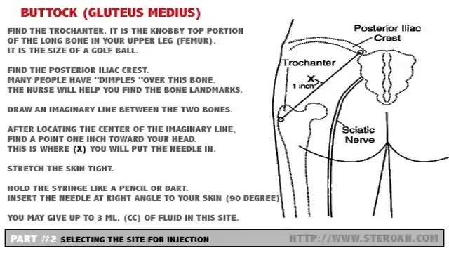
Carefully select the site for injection so major blood vessels and nerves are avoided. Buttock (Gluteus Medius)- Hip (Ventrogluteal) - Leg (Vastus Lateralis) - Arm (Deltoid)
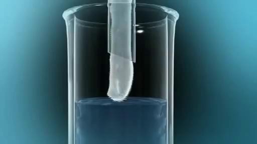
Intrauterine insemination (IUI) is a fertility treatment that involves placing sperm inside a woman's uterus to facilitate fertilization. The goal of IUI is to increase the number of sperm that reach the fallopian tubes and subsequently increase the chance of fertilization
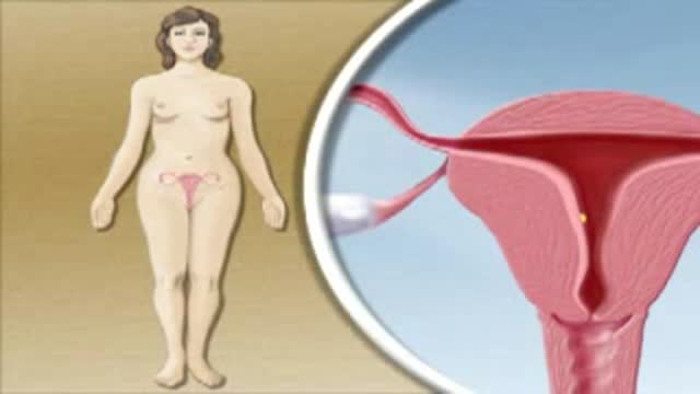
Ectopic Pregnancy
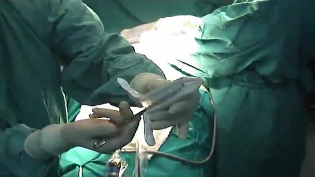
Internal hemorrhoids and loose rectal mucosa may block the exposure during the purse string suturing in stapled hemorrhooidopexy, and this may cause some complications. To retract the prolapsing rectal mucosa we modified the purse string anoscope of the PPH01 kit (Ethicon-Endosurgery, Cincinnati, O...H, USA) and produced a special anoscope. The open part of the purse string suture anoscope is covered by transparent acrylic (Orthoacryl�, Dentaurum, Pforzheim, Germany). The covering material had complete cylindrical outer and inner surfaces and was thin enough to let the anoscope easily rotate in the anal dilator and to let the 26 mm curved, round bodied needle of the 2/0 polypropilene suture move in the anoscope. A window, 3 cm long and 3-4 mm wide, was opened at the angled part of the anoscope 2 cm to the tip of the anoscope. This special anoscope was used for the purse string suture during stapled hemorrhoidopexy procedure in five patients. No postoperative complications, early or late, were encountered, and we propose that stapled hemorrhoidopexy procedure can be applied more easily by using this special anoscope.

A baby born with her heart pumping outside her body has stunned her parents and doctors in India.
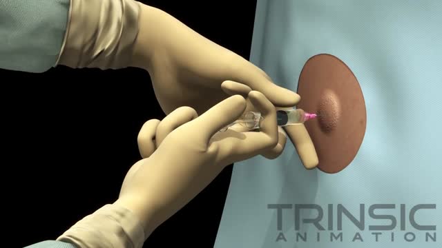
Thoracentesis (THOR-ah-sen-TE-sis) is a procedure to remove excess fluid in the space between the lungs and the chest wall. This space is called the pleural space. Normally, the pleural space is filled with a small amount of fluid—about 4 teaspoons full.

Kathleen Carr, MD performs a full shoulder exam for the musculoskeletal program at the University of Wisconsin Department of Family Medicine.
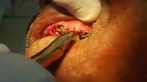
Watch that video of Gross Teeth Full of Worms Removal
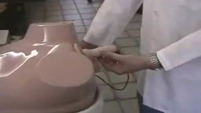
Bladder and prostate injection- Botox
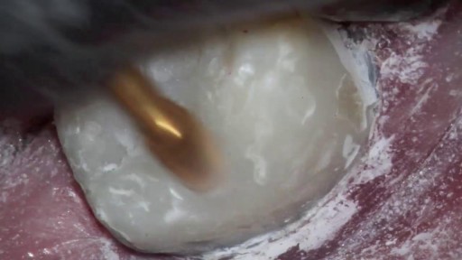
Watch that video of Filling Monster Tooth Cavity
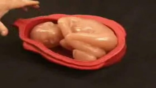
Leopold's Maneuvers are difficult to perform on obese women and women who have hydramnios. The palpation can sometimes be uncomfortable for the woman if care is not taken to ensure she is relaxed and adequately positioned. To aid in this, the health care provider should first ensure that the woman has recently emptied her bladder. If she has not, she may need to have a straight urinary catheter inserted to empy it if she is unable to micturate herself. The woman should lie on her back with her shoulders raised slightly on a pillow and her knees drawn up a little. Her abdomen should be uncovered, and most women appreciate it if the individual performing the maneuver warms their hands prior to palpation. First maneuver: Fundal Grip While facing the woman, palpate the woman's upper abdomen with both hands. A professional can often determine the size, consistency, shape, and mobility of the form that is felt. The fetal head is hard, firm, round, and moves independently of the trunk while the buttocks feel softer, are symmetric, and the shoulders and limbs have small bony processes; unlike the head, they move with the trunk. Second maneuver After the upper abdomen has been palpated and the form that is found is identified, the individual performing the maneuver attempts to determine the location of the fetal back. Still facing the woman, the health care provider palpates the abdomen with gentle but also deep pressure using the palm of the hands. First the right hand remains steady on one side of the abdomen while the left hand explores the right side of the woman's uterus. This is then repeated using the opposite side and hands. The fetal back will feel firm and smooth while fetal extremities (arms, legs, etc.) should feel like small irregularities and protrusions. The fetal back, once determined, should connect with the form found in the upper abdomen and also a mass in the maternal inlet, lower abdomen. Third maneuver: Pawlick's Grip In the third maneuver the health care provider attempts to determine what fetal part is lying above the inlet, or lower abdomen.[2] The individual performing the maneuver first grasps the lower portion of the abdomen just above the symphysis pubis with the thumb and fingers of the right hand. This maneuver should yield the opposite information and validate the findings of the first maneuver. If the woman enters labor, this is the part which will most likely come first in a vaginal birth. If it is the head and is not actively engaged in the birthing process, it may be gently pushed back and forth. The Pawlick's Grip, although still used by some obstetricians, is not recommended as it is more uncomfortable for the woman. Instead, a two-handed approach is favored by placing the fingers of both hands laterally on either side of the presenting part. Fourth maneuver The last maneuver requires that the health care provider face the woman's feet, as he or she will attempt to locate the fetus' brow. The fingers of both hands are moved gently down the sides of the uterus toward the pubis. The side where there is resistance to the descent of the fingers toward the pubis is greatest is where the brow is located. If the head of the fetus is well-flexed, it should be on the opposite side from the fetal back. If the fetal head is extended though, the occiput is instead felt and is located on the same side as the back. Cautions Leopold's maneuvers are intended to be performed by health care professionals, as they have received the training and instruction in how to perform them. That said, as long as care taken not to roughly or excessively disturb the fetus, there is no real reason it cannot be performed at home as an informational exercise. It is important to note that all findings are not truly diagnostic, and as such ultrasound is required to conclusively determine the fetal position.
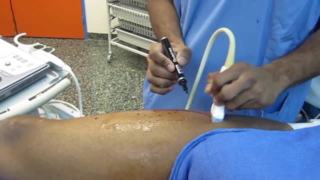
it's the video of the OR during a dynamic reconstruction of the achilles tendon by a composite anterolateral perforator flap
