
- Physical Examination
- Surgical Examination
- Ophthalmology
- Clinical Skills
- Orthopedics
- Surgery Videos
- Laparoscopy
- Pediatrics
- Funny Videos
- Cardiothoracic Surgery
- Nursing Videos
- Plastic Surgery
- Otorhinolaryngology
- Histology and Histopathology
- Neurosurgery
- Dermatology
- Pediatric Surgery
- Urology
- Dentistry
- Oncology and Cancers
- Anatomy Videos
- Health and Fitness
- Radiology
- Anaesthesia
- Physical Therapy
- Pharmacology
- Interventional Radiology
- Cardiology
- Endocrinology
- Gynecology
- Emergency Medicine
- Psychiatry and Psychology
- Childbirth Videos
- General Medical Videos
- Nephrology
- Physiology
- Diet and Food Health
- Diabetes Mellitus
- Neurology
- Women Health
- Osteoporosis
- Gastroenterology
- Pulmonology
- Hematology
- Rheumatology
- Toxicology
- Nuclear Medicine
- Infectious Diseases
- Vascular Disease
- Reproductive Health
- Burns and Wound Healing
- Kidney Zone Podcast
- Other

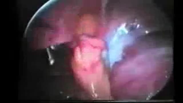
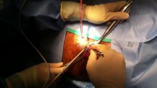

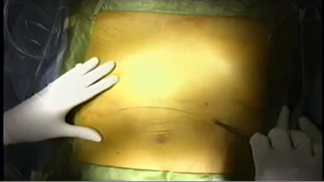
Whipple procedure

Educational only: videos on MedicalVideos.com are for general information and are not medical advice. Always consult your own doctor about your health.
What is a Whipple procedure?
Also called a pancreaticoduodenectomy, the Whipple procedure is performed to address chronic pancreatitis and cancer of the pancreas, ampulla of Vater, duodenum, and the distal bile duct. The Whipple procedure involves removing the cancerous parts of the pancreas, duodenum, common bile duct, and if required, part of the stomach.
This video in 30 seconds
A surgeon narrates a Whipple procedure performed for chronic pancreatitis. He talks through mobilising the pancreas, dissecting major veins, dividing the bile duct, duodenum, and pancreatic neck, removing the specimen, and then the reconstruction with pancreatic, biliary, and duodenal anastomoses, finishing with drains and a gastrostomy tube.
Key points
- The pancreas is mobilised and resectability is assessed at the start
- The superior mesenteric and portal veins are dissected during the procedure
- The bile duct, duodenum, and pancreatic neck are divided in sequence
- The specimen including the uncinate process is removed before reconstruction
- Reconstruction is described as pancreatic, biliary, and duodenal anastomoses
Chapters
- 0:02 — Indication and initial mobilisation
- 0:20 — Superior mesenteric vein dissection
- 0:45 — Bile duct division
- 1:09 — Pancreatic neck division
- 1:55 — Preparing for reconstruction
- 2:25 — Pancreatic anastomosis
- 3:55 — Biliary anastomosis
- 4:18 — Duodenal anastomosis
Video Transcript
A surgeon's running narration of a Whipple procedure performed for chronic pancreatitis. He talks through mobilizing the pancreas, dissecting the superior mesenteric and portal veins, dividing the bile duct, duodenum, and pancreatic neck, removing the specimen including the uncinate process, and the reconstruction — pancreatic, biliary, and duodenal anastomoses — finishing with drains and a gastrostomy tube.
Read the full transcript
Most whipple procedures are done for pancreas. Today we're operating for chronic pancreatitis, so we skipped the usual laparoscopy and proceeded directly to the midline incision. We initially mobilized the pancreas from the retroperitoneum and assess receptability. The next phase of the operation we proceed to dissecting the superior mesenteric vein, which is critical to assessing receptability. The vein is dissected out from beneath the neck of the pancreas and clearance made. We subsequently move to the hepatoduodenal ligament and identify the hepatic artery and gastroduodenal arteries. The gastroduodenal artery is divided,
which opens the access to the portal vein. At this point the bile duct is then divided, allowing further dissection down the portal vein. Once the hepatoduodenal ligament is completed, we divide the duodenum and proceed to divide the proximal jejunum. This now isolates the specimen to include only the connections to the pancreas. We divide the neck of the pancreas just over the portal vein. In this case a stent had been placed in the pancreatic duct, and that is divided in addition to the pancreas. The stent was maintaining patency of the pancreatic
duct. We separate the head of the pancreas from the portal vein, and then comes the most dangerous part of the operation. The unsonate process is immediately adjacent to the superior mesenteric artery and must be divided very carefully. There are large vessels in this tissue, despite the appearing innocence of the tissue. At this point the specimen is completely removed, and we can begin to prepare for the reconstruction. Now what remains is the superior mesenteric veins, planic vein, portal vein, and you can see the bile duct in the left upper corner. The superior
mesenteric artery is just below the specimen. Both ends are closed off by staple line, and the cut surface of the pancreas still has the stent. For the reconstruction we bring the duodenum through the transverse mesocolon and create a side to side pancreatic ogegenostomy as the first anastomosis. This is done in a dunked fashion using sutures in the outer layer placed well back on the pancreatic capsule. The inner layer of this dunked anastomosis is a fine absorbable material, and just the cut edge of pancreas is brought to the cut edge of
gejunal mucosa. As this anastomosis is completed, in the final stages of this anastomosis, the second layer is placed on the capsule of the pancreas, the gejunum is folded over to complete the dunking process. The next anastomosis is the biliary anastomosis. In this case we're dealing with a very small fine bile duct, so interrupted sutures of fine absorbable material are used to create the end to side colidocogegenostomy. Several centimeters downstream and end to side duodenugegenostomy is performed using a single layer absorbable running suture. To complete the operation I always place a gastrostomy tube for GI tract drainage and a single suction drainage
catheter adjacent to the colidocogegenostomy and pancreaticogegenostomy.
Transcript auto-generated from the video’s audio for accessibility and study. Educational content only — not medical advice.
Video Overview
From our Surgery Videos collection: Surgery videos: watch real operations step by step — general, laparoscopic and specialist surgery performed in the OR, explained for students and surgeons. Browse all Surgery Videos videos →
Medical Videos
Free medical and surgery videos for patients, students and professionals — trusted since 2007.
Founded by Dr. Mohamed Ibrahim, MD, PhD — physician-scientist.
Follow us on Facebook · Instagram
SORT BY-
Top Comments
-
Latest comments