- Physical Examination
- Surgical Examination
- Ophthalmology
- Clinical Skills
- Orthopedics
- Surgery Videos
- Laparoscopy
- Pediatrics
- Funny Videos
- Cardiothoracic Surgery
- Nursing Videos
- Plastic Surgery
- Otorhinolaryngology
- Histology and Histopathology
- Neurosurgery
- Dermatology
- Pediatric Surgery
- Urology
- Dentistry
- Oncology and Cancers
- Anatomy Videos
- Health and Fitness
- Radiology
- Anaesthesia
- Physical Therapy
- Pharmacology
- Interventional Radiology
- Cardiology
- Endocrinology
- Gynecology
- Emergency Medicine
- Psychiatry and Psychology
- Childbirth Videos
- General Medical Videos
- Nephrology
- Physiology
- Diet and Food Health
- Diabetes Mellitus
- Neurology
- Women Health
- Osteoporosis
- Gastroenterology
- Pulmonology
- Hematology
- Rheumatology
- Toxicology
- Nuclear Medicine
- Infectious Diseases
- Vascular Disease
- Reproductive Health
- Burns and Wound Healing
- Kidney Zone Podcast
- Other
Costal Cartilages
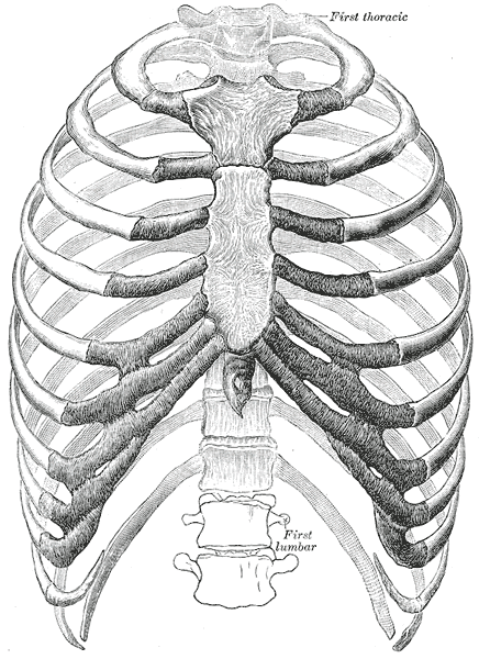
Gray's Anatomy (1918) plate, public domain
Costal cartilages are the bars of firm, flexible cartilage that connect the bony ribs to the sternum, or to the cartilage above. They are what make the front of the chest wall springy rather than rigid, letting the cage expand with breathing and absorb knocks. With age they gradually calcify and stiffen.
Key facts
- The first seven pairs join the sternum directly.
- Pairs 8 to 10 join the cartilage above, forming the costal margin.
- They are made of hyaline cartilage, like the covering of joint surfaces.
- Their elasticity returns the chest wall after each breath out.
- They contain no blood vessels, nourished instead by their surface membrane.
What can go wrong
The classic costal-cartilage problem is costochondritis — inflammation at the junctions with the sternum, producing localised chest pain that is tender to press. Because it mimics heart pain, new chest pain is always assessed medically first. The cartilages can also be strained by heavy coughing or injured in chest trauma.
Related procedures & tests
Related symptoms
Watch: related videos
Frequently asked questions
Why does the front of the rib cage use cartilage instead of bone?
Cartilage gives the chest wall spring. If the whole cage were rigid bone, breathing movement would be minimal and the cage would fracture more easily under the knocks it is designed to absorb.
What is costochondritis?
Inflammation where a rib's cartilage joins the breastbone. It causes a sharp or aching pain that is characteristically tender when pressed, though any new chest pain should first be checked to exclude heart causes.
Educational information only. This atlas explains normal anatomy and describes conditions in general terms; it is not medical advice and does not replace assessment by a qualified clinician. If you have symptoms that worry you, seek medical care.