- Physical Examination
- Surgical Examination
- Ophthalmology
- Clinical Skills
- Orthopedics
- Surgery Videos
- Laparoscopy
- Pediatrics
- Funny Videos
- Cardiothoracic Surgery
- Nursing Videos
- Plastic Surgery
- Otorhinolaryngology
- Histology and Histopathology
- Neurosurgery
- Dermatology
- Pediatric Surgery
- Urology
- Dentistry
- Oncology and Cancers
- Anatomy Videos
- Health and Fitness
- Radiology
- Anaesthesia
- Physical Therapy
- Pharmacology
- Interventional Radiology
- Cardiology
- Endocrinology
- Gynecology
- Emergency Medicine
- Psychiatry and Psychology
- Childbirth Videos
- General Medical Videos
- Nephrology
- Physiology
- Diet and Food Health
- Diabetes Mellitus
- Neurology
- Women Health
- Osteoporosis
- Gastroenterology
- Pulmonology
- Hematology
- Rheumatology
- Toxicology
- Nuclear Medicine
- Infectious Diseases
- Vascular Disease
- Reproductive Health
- Burns and Wound Healing
- Kidney Zone Podcast
- Other
Thoracic Cage
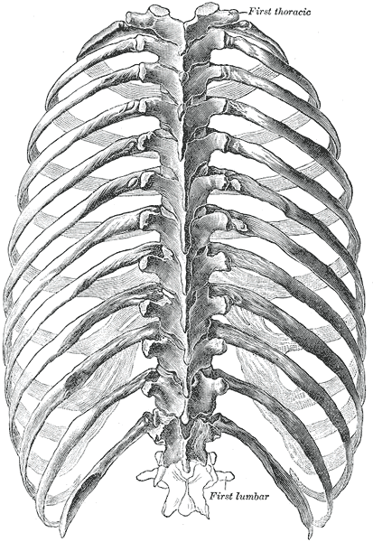
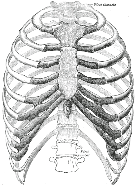
Gray's Anatomy (1918) plate, public domain
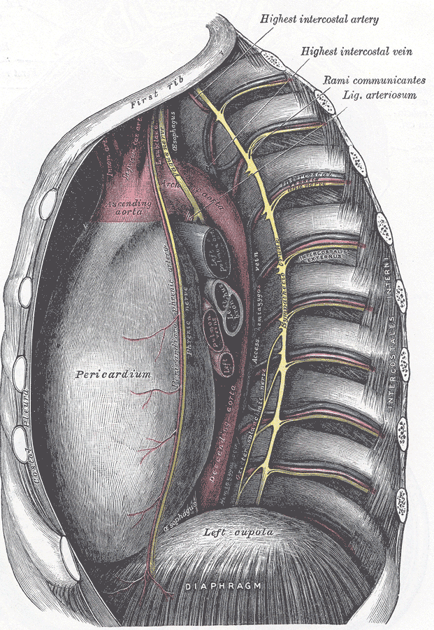
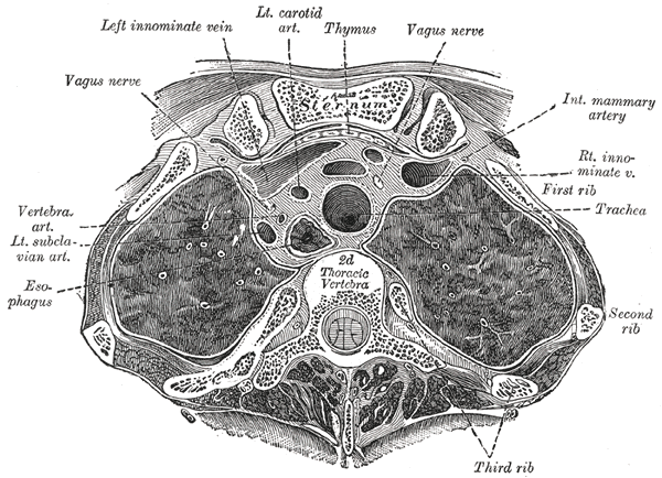
The thoracic cage — the rib cage — is the bony basket of the chest: 12 pairs of ribs, the sternum in front, the costal cartilages joining them, and the 12 thoracic vertebrae behind. It protects the heart, lungs and great vessels, supports the shoulder girdle, and expands and recoils with every breath.
Key facts
- The cage is formed by 24 ribs, the sternum, costal cartilages and 12 thoracic vertebrae.
- It protects the heart, lungs, liver, spleen and upper kidneys by position.
- Its shape is a barrel — wider below than above, and wider side-to-side than front-to-back.
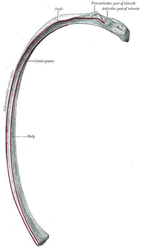
- The intercostal muscles between the ribs drive its breathing movements.
- Its lower rim (the costal margin) can be felt below the chest.
What can go wrong
Rib fractures are the commonest injury of the cage, usually from falls or blows; the main concern is injury to the lung or organs beneath rather than the rib itself. In older adults the cage stiffens as cartilages calcify, reducing chest expansion. A sunken or funnel-shaped front (pectus excavatum) is a developmental shape variation sometimes assessed in adolescence.
Related procedures & tests
Related symptoms
Related muscles
Frequently asked questions
Why is the rib cage springy rather than solid?
Breathing needs movement: the cage must expand thousands of times a day for a lifetime. Cartilage joints and angled ribs let it move like a bucket handle while still shielding the organs inside.
What is the weakest part of the rib cage?
The ribs themselves are weakest at their angle — the bend at the back — and the upper ribs are shielded by the shoulder, so fractures most often involve the middle ribs (about the 4th to 10th).
Educational information only. This atlas explains normal anatomy and describes conditions in general terms; it is not medical advice and does not replace assessment by a qualified clinician. If you have symptoms that worry you, seek medical care.