- Physical Examination
- Surgical Examination
- Ophthalmology
- Clinical Skills
- Orthopedics
- Surgery Videos
- Laparoscopy
- Pediatrics
- Funny Videos
- Cardiothoracic Surgery
- Nursing Videos
- Plastic Surgery
- Otorhinolaryngology
- Histology and Histopathology
- Neurosurgery
- Dermatology
- Pediatric Surgery
- Urology
- Dentistry
- Oncology and Cancers
- Anatomy Videos
- Health and Fitness
- Radiology
- Anaesthesia
- Physical Therapy
- Pharmacology
- Interventional Radiology
- Cardiology
- Endocrinology
- Gynecology
- Emergency Medicine
- Psychiatry and Psychology
- Childbirth Videos
- General Medical Videos
- Nephrology
- Physiology
- Diet and Food Health
- Diabetes Mellitus
- Neurology
- Women Health
- Osteoporosis
- Gastroenterology
- Pulmonology
- Hematology
- Rheumatology
- Toxicology
- Nuclear Medicine
- Infectious Diseases
- Vascular Disease
- Reproductive Health
- Burns and Wound Healing
- Kidney Zone Podcast
- Other
Pineal Gland
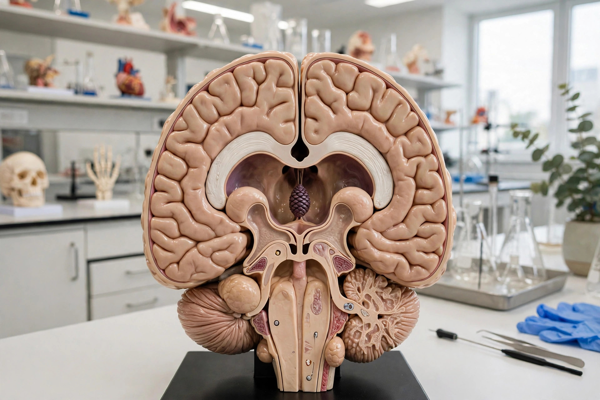
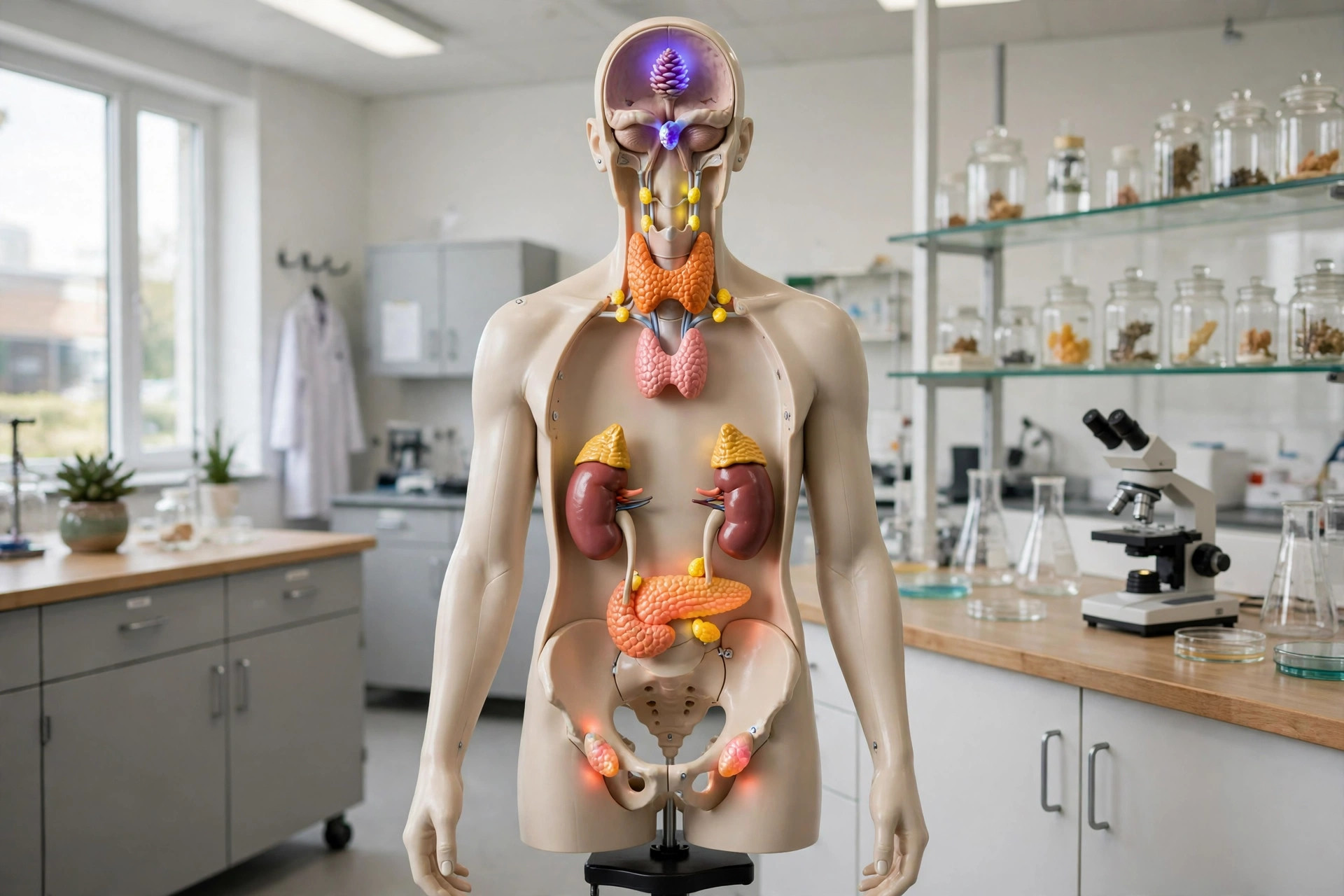
AI-generated anatomical illustration
The pineal gland is a tiny pinecone-shaped structure, about 8 mm long, sitting at the back of the third ventricle deep in the brain. It secretes melatonin in darkness: light signals from the eyes, relayed via the hypothalamus's clock nucleus, suppress it by day. Melatonin thereby signals night-time to the body and helps set sleep timing and seasonal rhythms.
Key facts
- It is one of the few unpaired midline structures in the brain.
- The gland often calcifies with age (brain sand), making it a useful midline landmark on scans.
- Melatonin secretion peaks in the small hours and falls before waking, aligning sleep with darkness.
- Unlike most of the brain, the pineal sits outside the blood-brain barrier, sampling blood chemistry directly.
What can go wrong
Pineal problems are uncommon: tumours of the region (in children and young adults) can block the fluid pathways of the brain, causing headache and raised pressure, or press on eye-movement centres. Cysts of the pineal are frequent incidental scan findings and are usually harmless. Melatonin supplements are widely used for jet lag and some sleep problems, though the gland itself rarely fails.
Related procedures & tests
Related symptoms
Frequently asked questions
What does melatonin do?
It is the hormone of darkness: rising levels in the evening promote sleepiness and help synchronise the body's daily rhythms with the light-dark cycle.
Are pineal cysts dangerous?
Almost always not. They are common incidental findings on MRI, usually small and stable, and are typically left alone unless they cause symptoms.
Educational information only. This atlas explains normal anatomy and describes conditions in general terms; it is not medical advice and does not replace assessment by a qualified clinician. If you have symptoms that worry you, seek medical care.