
- Physical Examination
- Surgical Examination
- Ophthalmology
- Clinical Skills
- Orthopedics
- Surgery Videos
- Laparoscopy
- Pediatrics
- Funny Videos
- Cardiothoracic Surgery
- Nursing Videos
- Plastic Surgery
- Otorhinolaryngology
- Histology and Histopathology
- Neurosurgery
- Dermatology
- Pediatric Surgery
- Urology
- Dentistry
- Oncology and Cancers
- Anatomy Videos
- Health and Fitness
- Radiology
- Anaesthesia
- Physical Therapy
- Pharmacology
- Interventional Radiology
- Cardiology
- Endocrinology
- Gynecology
- Emergency Medicine
- Psychiatry and Psychology
- Childbirth Videos
- General Medical Videos
- Nephrology
- Physiology
- Diet and Food Health
- Diabetes Mellitus
- Neurology
- Women Health
- Osteoporosis
- Gastroenterology
- Pulmonology
- Hematology
- Rheumatology
- Toxicology
- Nuclear Medicine
- Infectious Diseases
- Vascular Disease
- Reproductive Health
- Burns and Wound Healing
- Kidney Zone Podcast
- Other
Brain MRI
A brain MRI is the most detailed routine picture of the brain available, used for everything from stroke and tumour assessment to headache and seizure work-ups. This page explains what it shows, how it differs from a head CT, and what the report language means, in plain language with videos.
✓ Medically reviewed by Dr. Mohamed Ibrahim, MD, PhD — physician-scientist · Last reviewed: November 2026
Watch: Brain MRI videos
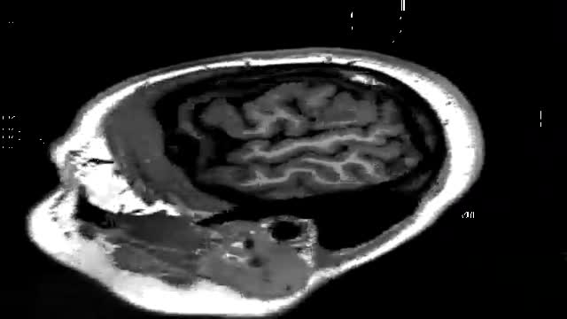 MRI of the brain13,599 views
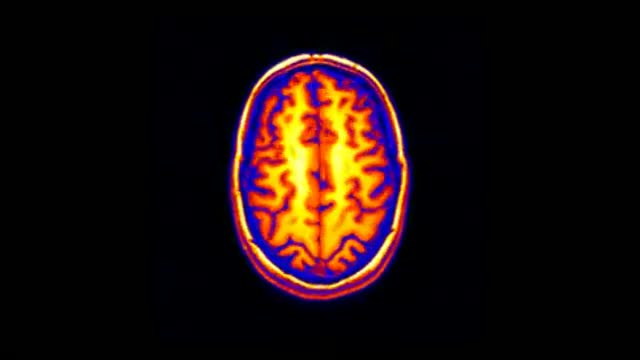 3D MRI of Human Brain9,124 views
 MRI of Fetal Brain Development8,098 views
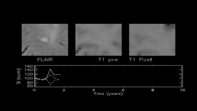 MRI Uses in Multiple Sclerosis4,430 views
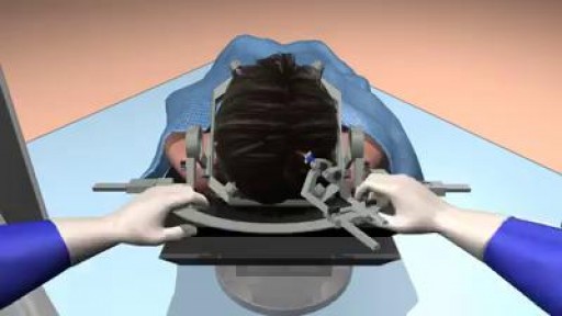 MRI-guided laser ablation for minimal invasive Neurosurgery. Kareem A Samer3,468 views
Guide
What it is
You lie on the scanner table with your head resting inside a padded frame that helps the machine pick up signals from the brain. The table slides into the scanner so your head is at its centre. A series of pictures is taken, each a few minutes long, with different settings to highlight different tissues — fluid, for example, looks bright on some sequences and dark on others, and comparing them is how radiologists characterise problems. Contrast dye through a vein is added for many brain examinations. The whole appointment usually takes well under an hour.
What it shows
Brain MRI shows the structure of the brain in fine detail: tumours, stroke damage (including early strokes CT can miss), bleeding, infection, and the inflammation plaques of multiple sclerosis. It examines the pituitary gland, the inner ear and hearing nerves, the eyes' pathways, and the blood vessels when angiography sequences are added. Special sequences can show the direction of fibre tracts or measure activity. It does not diagnose problems that leave no structural mark — many headaches, migraines, and functional symptoms have a normal MRI, which is itself useful information.
How to prepare
Preparation is the standard MRI safety screening — implants, metal fragments, and devices — plus removing metal from the head and neck. For brain MRI you lie with your head inside a close-fitting frame (a 'head coil') that helps capture the signal, which some people find the most claustrophobic part; a mirror is often fitted so you can see out. Contrast is given for many brain scans through a vein in the arm, partway through. There is no fasting for most brain MRI unless sedation is planned, and you can drive afterwards unless you had a sedative.
What the report words mean
Brain MRI reports are detailed. 'Lesion' means any abnormal area and covers many causes — inflammation, scarring, stroke damage, or tumour. 'White matter hyperintensities' are small bright patches on certain sequences, common with ageing and migraine, and often harmless. 'No acute infarct' means no new stroke was seen. 'Enhancement' after contrast suggests active inflammation or a tumour with leaky vessels. 'Atrophy' describes shrinkage, judged against age. 'Incidental finding' flags something unrelated, such as a small cyst, that may simply need monitoring.
Common questions
Why an MRI rather than a CT for the brain?
CT is fast and better for fresh bleeding, fractures, and emergencies, and it is easier for very unwell patients. MRI shows the brain tissue itself in much more detail — early stroke damage, small tumours, inflammation, and subtle structural problems — without radiation. The two tests answer different questions and are often used together.
What are 'white matter changes' on the report?
It means one or more tiny bright spots on certain sequences, seen very commonly as people age. They are usually linked to small changes in the tiny blood vessels of the brain and are often an incidental, harmless finding. Your clinician will judge whether they matter in your case, particularly if you have vascular risk factors.
Can a brain MRI diagnose multiple sclerosis?
It often can. MRI is one of the main tests used to support a diagnosis of multiple sclerosis because it shows the characteristic patches of inflammation and scarring (lesions) in the brain and spinal cord, including old and new ones side by side. The diagnosis also depends on symptoms, examination, and sometimes lumbar puncture results.
Can I move during the scan?
Mostly yes. You can blink and swallow normally, but try to keep your head still during each picture sequence, because movement blurs the images. You can rest, breathe, and shift slightly between sequences. If you feel unwell or panicky, squeeze the call button and the team can pause the scan.
More in MRI
Related
This guide is general information, not medical advice, and does not replace advice from your own clinician. How a scan is done, and whether contrast is used, depends on your own medical history — always follow the instructions of the team performing your scan, and tell them about kidney problems, allergies, implants, and the medicines you take. A normal brain MRI is reassuring, but some conditions — including many causes of headache — are diagnosed clinically and may not show on any scan.