
- Physical Examination
- Surgical Examination
- Ophthalmology
- Clinical Skills
- Orthopedics
- Surgery Videos
- Laparoscopy
- Pediatrics
- Funny Videos
- Cardiothoracic Surgery
- Nursing Videos
- Plastic Surgery
- Otorhinolaryngology
- Histology and Histopathology
- Neurosurgery
- Dermatology
- Pediatric Surgery
- Urology
- Dentistry
- Oncology and Cancers
- Anatomy Videos
- Health and Fitness
- Radiology
- Anaesthesia
- Physical Therapy
- Pharmacology
- Interventional Radiology
- Cardiology
- Endocrinology
- Gynecology
- Emergency Medicine
- Psychiatry and Psychology
- Childbirth Videos
- General Medical Videos
- Nephrology
- Physiology
- Diet and Food Health
- Diabetes Mellitus
- Neurology
- Women Health
- Osteoporosis
- Gastroenterology
- Pulmonology
- Hematology
- Rheumatology
- Toxicology
- Nuclear Medicine
- Infectious Diseases
- Vascular Disease
- Reproductive Health
- Burns and Wound Healing
- Kidney Zone Podcast
- Other
MRI Scan
An MRI scan makes detailed pictures of the body's soft tissues using a powerful magnet and radio waves — no X-ray radiation at all. This page explains how it works, who needs extra safety checks, and what having an MRI actually feels like, in plain language with videos.
✓ Medically reviewed by Dr. Mohamed Ibrahim, MD, PhD — physician-scientist · Last reviewed: November 2026
Watch: MRI Scan videos
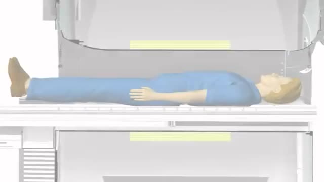 Understanding MRIs21,364 views
 Pediatric MRI, Kid-Friendly Service13,795 views
 Preparing Your Child for an MRI6,440 views
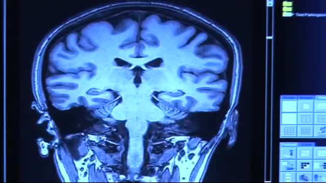 MRI Exam Procedure5,179 views
 Introduction to MRI Physics2,232 views
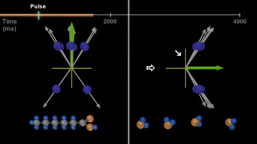 MRI: Basic Physics & a Brief History2,066 views
Guide
What it is
You lie on a table that slides into a tube-shaped scanner and stay as still as you can while the machine takes a series of pictures, each lasting a few minutes. The scanner makes loud tapping, buzzing, and banging noises as its magnets switch — you will be given earplugs or headphones, sometimes with music. The radiographer can see and hear you throughout and gives you a call button to squeeze if you need help. Depending on the area and the question, a whole examination commonly takes somewhere between about a quarter of an hour and an hour.
What it shows
MRI's strength is soft-tissue detail. It shows the brain and spinal cord, ligaments and cartilage in joints, muscles, and the organs of the pelvis better than any other scan, and it can characterise tumours, inflammation, and infection in fine detail. Special sequences highlight particular tissues or fluids, and a gadolinium-based contrast injection is sometimes added to make inflammation, tumours, and blood vessels stand out. MRI does not show cortical bone or air-filled lung as well as CT, and it is slower, so it is not the first test for most emergencies.
How to prepare
Safety screening is the main preparation. Before the scan you will complete a questionnaire about pacemakers and other implants, heart or brain surgery, metal fragments — especially in the eyes — and tattoos or permanent make-up, because some metal can move or heat in the magnet. Most modern implants are MRI-safe or safe under conditions, but the team must know exactly what you have. You will remove all loose metal, jewellery, cards, and hearing aids, and change into a gown. Tell the team if you are claustrophobic, pregnant, or have kidney problems (relevant if contrast is planned).
What the report words mean
MRI reports describe signal — brightness — rather than density. 'High signal' areas are bright and 'low signal' areas dark on a given sequence, and the pattern across sequences is what identifies tissue types. A 'lesion' is any abnormal spot, not necessarily a tumour. 'Enhancement' after contrast means tissue takes up the dye, suggesting active inflammation, tumour, or scar. Reports often compare with previous scans, so 'unchanged' or 'stable' is usually reassuring. The conclusion section summarises what the radiologist thinks the findings mean and whether follow-up is advised.
Common questions
Does MRI use radiation?
No. MRI uses a strong magnetic field and radio waves, not X-rays or ionising radiation, so there is no radiation dose from the scan itself. This is one reason MRI is often preferred for younger patients and for scans that need repeating, when it can answer the clinical question.
Can I have an MRI with a pacemaker or metal implant?
Often yes, but it depends on the exact device. Many modern pacemakers and implants are designed to be safe in MRI under set conditions, while a few older devices are not. This is why the team asks detailed questions and may check your implant records or contact the clinic that fitted it before scanning you.
Why is MRI so noisy, and can I move?
The banging comes from the magnet's gradient coils switching on and off rapidly — it is normal and expected, and hearing protection is always provided. Movement blurs the pictures in the sequence being taken, so you will be asked to keep the scanned area still; you can usually shift and get comfortable between sequences.
What if I am claustrophobic?
Tell the booking team in advance. Options include a wider or shorter scanner where available, a mirror or prism glasses to see out, a relative staying in the room (after screening), and, if needed, a mild sedative arranged through your clinician — in which case you cannot drive afterwards. Many anxious patients complete the scan successfully with these measures.
More in MRI
Related
Sources
This guide is general information, not medical advice, and does not replace advice from your own clinician. How a scan is done, and whether contrast is used, depends on your own medical history — always follow the instructions of the team performing your scan, and tell them about kidney problems, allergies, implants, and the medicines you take. Never assume an implant or device is MRI-safe — always tell the imaging team exactly what you have, even if a previous scan went ahead.