
- Physical Examination
- Surgical Examination
- Ophthalmology
- Clinical Skills
- Orthopedics
- Surgery Videos
- Laparoscopy
- Pediatrics
- Funny Videos
- Cardiothoracic Surgery
- Nursing Videos
- Plastic Surgery
- Otorhinolaryngology
- Histology and Histopathology
- Neurosurgery
- Dermatology
- Pediatric Surgery
- Urology
- Dentistry
- Oncology and Cancers
- Anatomy Videos
- Health and Fitness
- Radiology
- Anaesthesia
- Physical Therapy
- Pharmacology
- Interventional Radiology
- Cardiology
- Endocrinology
- Gynecology
- Emergency Medicine
- Psychiatry and Psychology
- Childbirth Videos
- General Medical Videos
- Nephrology
- Physiology
- Diet and Food Health
- Diabetes Mellitus
- Neurology
- Women Health
- Osteoporosis
- Gastroenterology
- Pulmonology
- Hematology
- Rheumatology
- Toxicology
- Nuclear Medicine
- Infectious Diseases
- Vascular Disease
- Reproductive Health
- Burns and Wound Healing
- Kidney Zone Podcast
- Other
Knee MRI
A knee MRI is the standard detailed scan for knee injuries and persistent knee pain, showing the cartilage, ligaments, and menisci that X-rays cannot. This page explains when it is used, what it finds, and how to read the report, in plain language with videos.
✓ Medically reviewed by Dr. Mohamed Ibrahim, MD, PhD — physician-scientist · Last reviewed: November 2026
Watch: Knee MRI videos
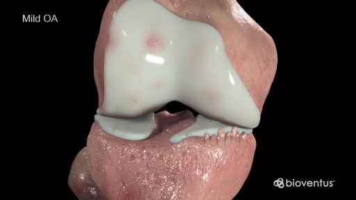 Stages of Knee Osteoarthritis17,375 views
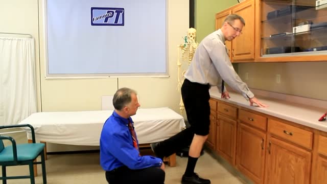 Meniscus Tear2,155 views
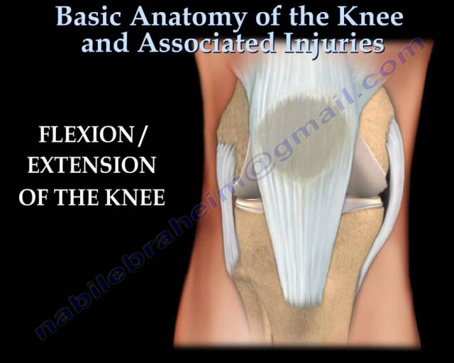 Knee injury ,Injuries - Everything You Need To Know - Dr. Nabil Ebraheim127 views
Guide
What it is
You lie on the scanner table with your leg extended and supported inside a special coil, and the table slides in until your knee is at the centre of the magnet — your head remains outside the tube. A series of pictures is taken in different planes (side, front, and cross-section) and with different settings to highlight cartilage, fluid, and ligaments in turn. The examination typically takes around twenty to thirty minutes. Keeping the leg still is essential, because even small movements blur the fine detail the scan is ordered for.
What it shows
Knee MRI shows the menisci (the cartilage pads), the cruciate and collateral ligaments, tendons, joint cartilage, and the bone marrow — everything a plain X-ray cannot. It is the standard test for a suspected meniscus tear or ligament injury, unexplained knee pain or swelling, a knee that locks or gives way, and for planning surgery. It also shows bone bruising, early arthritis change, cysts, and tumours. It does not replace a careful examination, and it is less useful for problems that are mainly about how the knee moves, which are assessed clinically.
How to prepare
Preparation follows the standard MRI safety screening for implants and metal. For a knee scan, only your leg goes into the centre of the scanner — your head and most of your body stay outside, so most people do not find it claustrophobic. You will wear shorts or a gown so the knee can be positioned, sometimes in a close-fitting coil that supports it. The key requirement is keeping the leg completely still; cushions help. Contrast dye is not usually needed for knee problems, though it is sometimes added for infection or tumour questions.
What the report words mean
Knee MRI reports go structure by structure. A 'meniscal tear' is graded and located — horizontal tears are usually wear-related, while displaced or 'bucket-handle' tears can lock the knee. 'Chondral loss' or 'chondropathy' means cartilage thinning; 'full-thickness' means worn down to bone. 'Bone marrow oedema' is bruising-like change inside the bone, often from injury or overload. An 'effusion' is extra fluid in the joint. 'Intact cruciate and collateral ligaments' is the reassuring phrase for the main stabilising ligaments. Mild degenerative wording is extremely common in pain-free knees, so reports are judged alongside symptoms.
Common questions
Why an MRI and not just an X-ray?
An X-ray shows bones and joint space only, so it is the usual first test for arthritis and fractures. MRI shows the soft tissues the X-ray cannot — cartilage, menisci, ligaments, tendons, and the bone marrow — so it is used when the X-ray does not explain the symptoms, or when a soft-tissue injury is suspected.
The report says I have a meniscus tear. Do I need surgery?
Quite often, no. Many meniscus tears — especially wear-and-tear tears in middle-aged knees — settle with physiotherapy and time, and studies show surgery is no better for many of them. Surgery is more likely to help if the knee locks, gives way, or a torn fragment is jamming the joint. Your clinician will weigh the scan against your symptoms.
Is MRI as good as keyhole surgery (arthroscopy) for diagnosis?
Arthroscopy (keyhole surgery with a camera) shows the inside of the joint directly and can treat problems at the same time, but it is an operation with anaesthetic and recovery time. MRI gives most of the same diagnostic information without surgery, so it is normally used first to decide whether arthroscopy is worthwhile.
Will I need a contrast injection?
Not usually. A knee MRI is done without contrast in most cases. Contrast is added for specific questions, such as infection, tumours, or assessing a joint after previous surgery. If it is planned, it is given through a vein in the arm partway through the scan.
More in MRI
Related
This guide is general information, not medical advice, and does not replace advice from your own clinician. How a scan is done, and whether contrast is used, depends on your own medical history — always follow the instructions of the team performing your scan, and tell them about kidney problems, allergies, implants, and the medicines you take. Many knee MRI findings, such as small meniscus tears, also occur in people with no pain at all — treatment decisions should be based on symptoms as well as the scan.