
- Physical Examination
- Surgical Examination
- Ophthalmology
- Clinical Skills
- Orthopedics
- Surgery Videos
- Laparoscopy
- Pediatrics
- Funny Videos
- Cardiothoracic Surgery
- Nursing Videos
- Plastic Surgery
- Otorhinolaryngology
- Histology and Histopathology
- Neurosurgery
- Dermatology
- Pediatric Surgery
- Urology
- Dentistry
- Oncology and Cancers
- Anatomy Videos
- Health and Fitness
- Radiology
- Anaesthesia
- Physical Therapy
- Pharmacology
- Interventional Radiology
- Cardiology
- Endocrinology
- Gynecology
- Emergency Medicine
- Psychiatry and Psychology
- Childbirth Videos
- General Medical Videos
- Nephrology
- Physiology
- Diet and Food Health
- Diabetes Mellitus
- Neurology
- Women Health
- Osteoporosis
- Gastroenterology
- Pulmonology
- Hematology
- Rheumatology
- Toxicology
- Nuclear Medicine
- Infectious Diseases
- Vascular Disease
- Reproductive Health
- Burns and Wound Healing
- Kidney Zone Podcast
- Other
CT Scan
A CT scan uses X-rays taken from many angles to build detailed cross-section pictures of the body — far more detailed than a plain X-ray. This page explains how CT works, when a contrast dye is added, and what happens during the scan, in plain language with videos.
✓ Medically reviewed by Dr. Mohamed Ibrahim, MD, PhD — physician-scientist · Last reviewed: November 2026
Watch: CT Scan videos
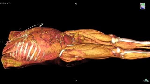 Whole Body CT scan with contrast media HD31,086 views
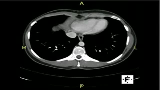 CT scan Abdomen26,633 views
 CT Scan -- Chest19,892 views
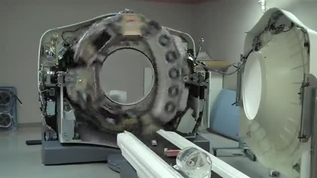 CT Scanner 64 slice Inside HD17,463 views
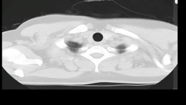 CT scan chest16,698 views
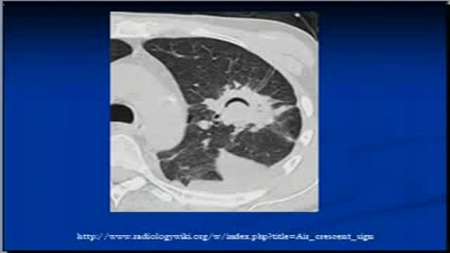 Chest x-ray -- CT Scan, air crescent sign15,375 views
Guide
What it is
You lie on a flat table that slides through a wide, open ring — CT is not a tunnel, and most people do not find it claustrophobic. Inside the ring, an X-ray source and detectors rotate around you, collecting measurements that a computer turns into thin slices. The table moves in small steps (or continuously, in modern spiral scanners) until the whole area is covered. The radiographer operates the scanner from the next room, watches you through a window, and can talk to you over an intercom. Each individual scan run takes only seconds to a minute or so.
What it shows
CT gives a detailed map of bones, organs, and blood vessels in almost any body part. It is the workhorse test for emergencies — head injuries, stroke assessment, chest pain, severe abdominal pain, and major trauma — and is widely used to investigate suspected cancer, plan surgery, and guide biopsies and drainage procedures. It shows the lungs, liver, bowel, and bones in far more detail than plain X-rays. Its limits are soft-tissue contrast in some areas, where MRI is better, and very early or purely functional problems that leave no structural mark.
How to prepare
Preparation depends on the body part. For many abdominal scans you are asked not to eat for a few hours beforehand, and you may drink a contrast liquid or have contrast through a vein. You will remove metal objects and wear a gown. Tell the team about kidney problems, previous reactions to contrast dye, asthma, thyroid problems, diabetes medicines such as metformin, and any chance of pregnancy — these affect how the scan is planned. If contrast is used, a small tube (cannula) is placed in your arm first, and drinking water afterwards helps your kidneys clear the dye.
What the report words mean
CT reports describe each organ in turn and end with a conclusion or impression — read that section first. A 'lesion' or 'mass' is any spot that looks different from the surrounding tissue; many are benign, and the word alone does not mean cancer. 'Enhancement' means tissue took up contrast dye, which tells the radiologist about its blood supply. 'Lymphadenopathy' means enlarged lymph nodes. 'No acute abnormality' means nothing needing urgent action was seen. Measurements are given in millimetres or centimetres so changes can be tracked between scans over time.
Common questions
Does a CT scan use a lot of radiation?
CT uses more radiation than a plain X-ray because it takes many X-ray measurements to build its detailed pictures. Scanners and protocols are designed to keep the dose as low as possible for the question being asked, and a needed CT is considered worthwhile when the result will change your care. If you are concerned, ask the team why the scan is recommended.
What does the contrast dye feel like?
When contrast is injected you may feel a warm flush spreading through your body, a metallic taste, and a brief sensation that feels like needing to pass urine — these are normal and fade within a minute or so. Tell the radiographer straight away if you feel itchy, sick, tight in the chest, or unwell in any way.
Can I have a CT if I am claustrophobic?
Most people with claustrophobia manage CT well because the scanner is a wide, open ring rather than a tunnel, and only the part being scanned is inside it. Your head often stays outside for body scans. Tell the team beforehand if you are anxious — they can talk you through it and keep the scan as short as possible.
Why might I need an MRI instead of a CT?
MRI shows some soft tissues — the brain, spinal cord, joints, and pelvic organs — in better detail than CT and uses no X-ray radiation. CT is faster, better for emergencies, the lungs, and bones, and is used when MRI is unavailable or unsuitable, for example with some metal implants. The ordering clinician chooses based on the question being asked.
More in CT
Related
Sources
- CT scan (MedlinePlus Medical Encyclopedia)
- CT scan (NHS)
- Computed Tomography (CT) (US Food & Drug Administration)
- Abdominal and Pelvic CT (RadiologyInfo.org (RSNA/ACR))
This guide is general information, not medical advice, and does not replace advice from your own clinician. How a scan is done, and whether contrast is used, depends on your own medical history — always follow the instructions of the team performing your scan, and tell them about kidney problems, allergies, implants, and the medicines you take. If you have kidney problems or have reacted to contrast dye before, tell the imaging team before the scan so they can plan safely.