
- Physical Examination
- Surgical Examination
- Ophthalmology
- Clinical Skills
- Orthopedics
- Surgery Videos
- Laparoscopy
- Pediatrics
- Funny Videos
- Cardiothoracic Surgery
- Nursing Videos
- Plastic Surgery
- Otorhinolaryngology
- Histology and Histopathology
- Neurosurgery
- Dermatology
- Pediatric Surgery
- Urology
- Dentistry
- Oncology and Cancers
- Anatomy Videos
- Health and Fitness
- Radiology
- Anaesthesia
- Physical Therapy
- Pharmacology
- Interventional Radiology
- Cardiology
- Endocrinology
- Gynecology
- Emergency Medicine
- Psychiatry and Psychology
- Childbirth Videos
- General Medical Videos
- Nephrology
- Physiology
- Diet and Food Health
- Diabetes Mellitus
- Neurology
- Women Health
- Osteoporosis
- Gastroenterology
- Pulmonology
- Hematology
- Rheumatology
- Toxicology
- Nuclear Medicine
- Infectious Diseases
- Vascular Disease
- Reproductive Health
- Burns and Wound Healing
- Kidney Zone Podcast
- Other
X-ray Basics
An X-ray is a quick, painless picture of the inside of the body made with a small, controlled burst of radiation. This page explains how X-rays work, what they can and cannot show, and how the radiation involved compares with everyday life, in plain language with videos.
✓ Medically reviewed by Dr. Mohamed Ibrahim, MD, PhD — physician-scientist · Last reviewed: November 2026
Watch: X-ray Basics videos
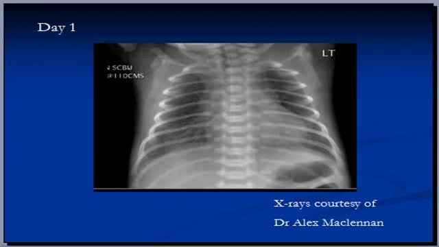 Chest x-ray interpretation, RDS40,674 views
 Chest x-ray, mediastinal structures39,485 views
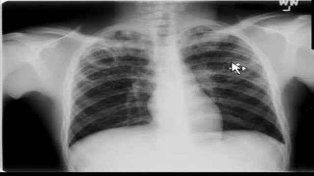 Chest x-ray --Tuberculosis30,298 views
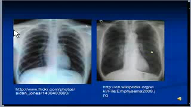 Chest x-ray interpretation -- COPD and Emphysema27,260 views
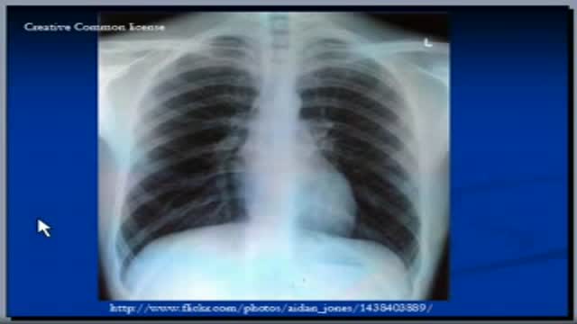 Chest x-ray interpretation, Lateral view25,896 views
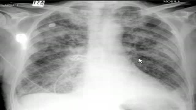 chest x-ray, pulmonary edema23,354 views
Guide
What it is
An X-ray machine sends a brief burst of invisible radiation through the part of the body being examined, onto a detector on the other side. The detector records how much radiation got through at each point, and a computer turns that into a flat, two-dimensional picture. The test is painless and usually takes only a few minutes, most of it spent positioning you correctly. A related technique, fluoroscopy, uses the same principle as a live moving picture, so doctors can watch a joint move, follow swallowed contrast through the gut, or guide a procedure in real time.
What it shows
X-rays are strongest where density differences are large. Broken or dislocated bones, joint wear, and some dental problems show up clearly. In the chest, X-rays show the air-filled lungs, the heart outline, and the ribs. They can reveal obvious problems such as a badly collapsed lung, a large build-up of fluid, or a dense pneumonia patch. X-rays are much weaker for soft tissues: muscles, ligaments, the brain, and early or subtle disease often look normal on an X-ray even when something is wrong, which is why a normal X-ray does not rule out every problem.
How to prepare
Most X-rays need no preparation at all. You may be asked to remove jewellery, watches, glasses, or clothing with metal fastenings over the area, because metal shows up bright white and can hide detail. You will usually wear a gown and be positioned by a radiographer, who may place the detector behind or under the body part. Staying still for a few seconds matters, because movement blurs the picture. Tell the team if there is any chance you could be pregnant, so they can decide with you whether the test should go ahead, be changed, or be postponed.
What the report words mean
Reports describe position and shading rather than naming diseases directly. An 'opacity' or 'shadow' is an area that looks whiter than expected — it means something denser than air is there, which has many possible causes. 'Lucency' means a darker area, where more X-rays passed through. 'Fracture' is a break, and 'alignment' describes whether bones sit in their normal position. 'No acute bony injury' means no fresh break was seen. The report is written for the clinician who ordered the test, who combines it with your symptoms and examination before deciding what it means.
Common questions
How much radiation does an X-ray use?
A single X-ray uses a very small amount of radiation — far less than a CT scan — and the dose is kept as low as reasonably possible while still giving a clear picture. The benefit of a needed X-ray is generally considered to outweigh this small risk, but X-rays are still only ordered when the result is expected to help your care.
Does an X-ray hurt?
No. You feel nothing from the X-rays themselves. The only discomfort is sometimes in holding a position for a few seconds, which can be awkward if the area is already injured or sore. Tell the radiographer if a position is too painful and they will adapt it where they can.
Why did I need more than one picture?
A single X-ray is a flat view from one direction, so a second view from another angle is often taken to show depth and to avoid missing a break or abnormality hidden behind another structure. Two or more views of the same area are routine, not a sign that something serious was found.
Can an X-ray show muscles, ligaments, or the brain?
Not well. X-rays show soft tissues only as vague grey shapes, so ligament tears, muscle injuries, and brain problems are usually assessed with ultrasound, MRI, or CT instead. An X-ray is often still the first test for an injured limb because it quickly rules a fracture in or out.
More in X-ray & Fluoroscopy
Related
Sources
- X-ray (MedlinePlus Medical Encyclopedia)
- X-ray (NHS)
- Medical X-ray Imaging (US Food & Drug Administration)
- Bone X-ray (RadiologyInfo.org (RSNA/ACR))
This guide is general information, not medical advice, and does not replace advice from your own clinician. How a scan is done, and whether contrast is used, depends on your own medical history — always follow the instructions of the team performing your scan, and tell them about kidney problems, allergies, implants, and the medicines you take. If you are pregnant or might be, always tell the imaging team before any X-ray, CT, or fluoroscopy examination.