
- Physical Examination
- Surgical Examination
- Ophthalmology
- Clinical Skills
- Orthopedics
- Surgery Videos
- Laparoscopy
- Pediatrics
- Funny Videos
- Cardiothoracic Surgery
- Nursing Videos
- Plastic Surgery
- Otorhinolaryngology
- Histology and Histopathology
- Neurosurgery
- Dermatology
- Pediatric Surgery
- Urology
- Dentistry
- Oncology and Cancers
- Anatomy Videos
- Health and Fitness
- Radiology
- Anaesthesia
- Physical Therapy
- Pharmacology
- Interventional Radiology
- Cardiology
- Endocrinology
- Gynecology
- Emergency Medicine
- Psychiatry and Psychology
- Childbirth Videos
- General Medical Videos
- Nephrology
- Physiology
- Diet and Food Health
- Diabetes Mellitus
- Neurology
- Women Health
- Osteoporosis
- Gastroenterology
- Pulmonology
- Hematology
- Rheumatology
- Toxicology
- Nuclear Medicine
- Infectious Diseases
- Vascular Disease
- Reproductive Health
- Burns and Wound Healing
- Kidney Zone Podcast
- Other
Ultrasound
An ultrasound scan makes live pictures of the body's insides using sound waves — no radiation, no injections, and no recovery time. This page explains how ultrasound works, what it is used for, and its limits, in plain language with videos.
✓ Medically reviewed by Dr. Mohamed Ibrahim, MD, PhD — physician-scientist · Last reviewed: November 2026
Watch: Ultrasound videos
 Vaginal Ultrasound89,034 views
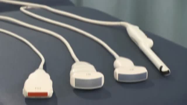 Ultrasound of Male Reproductive Organs63,146 views
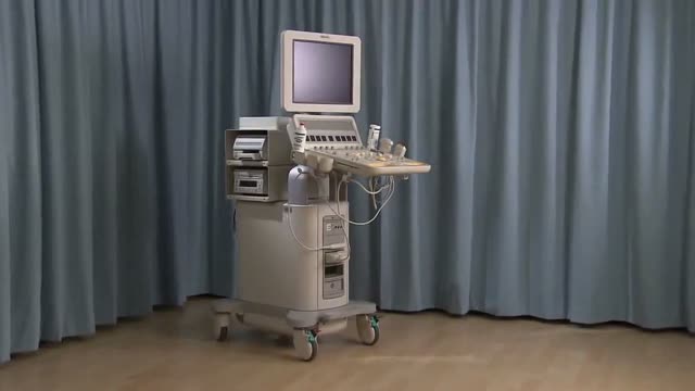 Ultrasound of the Breast25,264 views
 How to use Ultrasound in Pregnancy21,768 views
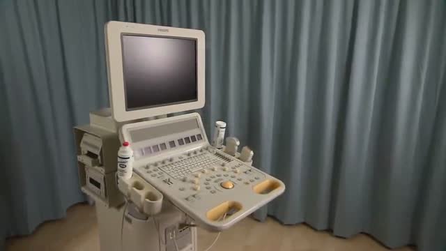 Ultrasound of the Thyroid and Parathyroid Glands17,785 views
 The Smallest Ultrasound device in the world10,624 views
Guide
What it is
During a scan, a sonographer (or a doctor) presses a small handheld probe against the skin with a layer of gel that helps the sound waves pass into the body. The probe is moved and angled to build up views of the organ from several directions, and still pictures and measurements are recorded. You may be asked to change position or hold your breath. Most scans take somewhere between fifteen and forty-five minutes depending on the area, and the probe pressure can be firm but should not be painful.
What it shows
Ultrasound is used across the body: the abdomen (liver, gallbladder, kidneys), the pelvis and pregnancy, the heart (as an echocardiogram), blood vessels (with Doppler), the thyroid, breasts, testicles, muscles and joints, and the eyes. It also guides procedures — biopsies, drainages, and injections — in real time. Its limits are physical: sound cannot pass through gas or bone, so the bowel, lungs, and anything hidden behind them are poorly seen, and image quality falls with depth in larger patients. CT or MRI is used when ultrasound cannot answer the question.
How to prepare
Preparation depends on the area. Abdominal scans usually ask for fasting; pelvic scans often need a full bladder; most other scans need nothing at all. You will expose the area being scanned and lie on a couch. The gel can feel cold at first. Internal scans — through the vagina or rectum — are offered for some pelvic questions because they give a closer, clearer view; they are done with a slim covered probe, a chaperone is available, and you can decline. The scan is painless apart from pressure, and there is no recovery time.
What the report words mean
Ultrasound reports describe echoes. Structures are 'hyperechoic' (bright), 'hypoechoic' (darker than surroundings), or 'anechoic' (black — usually simple fluid, like a cyst). 'Heterogeneous' means mixed in texture. Measurements are given in millimetres or centimetres and compared with previous scans. 'Limited study' means something — often bowel gas or body habitus — restricted the view, so conclusions are cautious. A 'sonographic appearance consistent with…' phrase means the picture fits a condition but the scan alone does not prove it; the ordering clinician completes the picture.
Common questions
Does ultrasound use radiation?
No. Ultrasound uses sound waves, not X-rays or ionising radiation, and no harmful effects have been shown at the power levels used for diagnostic scanning. This is why it is the standard imaging method throughout pregnancy and can be repeated safely whenever needed.
Why does the person doing the scan matter so much?
Ultrasound pictures are built from echoes returning to the probe, and the sonographer interprets them live, choosing what to measure and record. Experience matters more than with most scans, and some things can be missed or be hard to see — for example, if bowel gas blocks the view. Reports note when a scan was limited.
What is the difference between 2D, 3D, and 4D ultrasound?
Three-dimensional (3D) ultrasound assembles many 2D slices into a volume that can be viewed as a surface picture — most familiar from pregnancy scans. Four-dimensional (4D) is 3D shown moving in real time. They are additions to, not replacements for, the standard 2D examination that checks anatomy and measurements.
What is point-of-care ultrasound (POCUS)?
It is a small handheld ultrasound machine that connects to a tablet or screen. Clinicians use it at the bedside or in clinic to answer focused questions quickly — checking for fluid, guiding injections and drips, or a first look at the heart or bladder. A formal department scan is still needed for a complete, reported examination.
More in Ultrasound
Related
Sources
- Ultrasound (MedlinePlus Medical Encyclopedia)
- Ultrasound scan (NHS)
- General Ultrasound (RadiologyInfo.org (RSNA/ACR))
This guide is general information, not medical advice, and does not replace advice from your own clinician. How a scan is done, and whether contrast is used, depends on your own medical history — always follow the instructions of the team performing your scan, and tell them about kidney problems, allergies, implants, and the medicines you take. Ultrasound pictures depend heavily on the operator and the patient's body; a limited scan may need repeating or following up with another test.