
- Physical Examination
- Surgical Examination
- Ophthalmology
- Clinical Skills
- Orthopedics
- Surgery Videos
- Laparoscopy
- Pediatrics
- Funny Videos
- Cardiothoracic Surgery
- Nursing Videos
- Plastic Surgery
- Otorhinolaryngology
- Histology and Histopathology
- Neurosurgery
- Dermatology
- Pediatric Surgery
- Urology
- Dentistry
- Oncology and Cancers
- Anatomy Videos
- Health and Fitness
- Radiology
- Anaesthesia
- Physical Therapy
- Pharmacology
- Interventional Radiology
- Cardiology
- Endocrinology
- Gynecology
- Emergency Medicine
- Psychiatry and Psychology
- Childbirth Videos
- General Medical Videos
- Nephrology
- Physiology
- Diet and Food Health
- Diabetes Mellitus
- Neurology
- Women Health
- Osteoporosis
- Gastroenterology
- Pulmonology
- Hematology
- Rheumatology
- Toxicology
- Nuclear Medicine
- Infectious Diseases
- Vascular Disease
- Reproductive Health
- Burns and Wound Healing
- Kidney Zone Podcast
- Other
Echocardiogram
An echocardiogram — often just called an 'echo' — is an ultrasound scan of the heart that shows it beating, pumping, and leaking or narrowing at the valves. This page explains the types of echo, what the measurements mean, and how it links with ECG testing, in plain language with videos.
✓ Medically reviewed by Dr. Mohamed Ibrahim, MD, PhD — physician-scientist · Last reviewed: November 2026
Watch: Echocardiogram videos
 Echocardiography showing an Acute Pulmonary Embolism22,642 views
 Transthoracic Echocardiogram General6,822 views
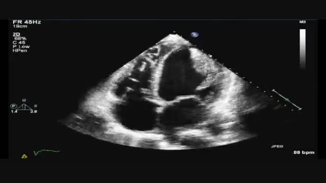 Transesophageal Echocardiogram of a Giant Thrombus in the Left Ventricle6,595 views
 Basic Transthoracic Echocardiogram5,840 views
Guide
What it is
In the standard test (a transthoracic echo) you lie on your side while a sonographer moves a probe over the chest with gel, taking pictures from several positions. Doppler is used throughout to measure the direction and speed of blood flow through the valves and chambers, shown in colour and as audible signals. The test usually takes around half an hour to an hour, is painless apart from firm probe pressure, and you can get up and carry on with your day straight away. Recordings are measured and reported afterwards by a specialist.
What it shows
An echocardiogram shows the size and shape of the four chambers, how thick the heart muscle is, and how strongly it squeezes. It shows all four valves opening, closing, and leaking, and it measures pressures and flow speeds using Doppler. It can reveal fluid around the heart, congenital heart problems, areas of muscle damaged by a heart attack, and signs of strain from high blood pressure or lung disease. It does not show the coronary arteries themselves in detail — narrowed heart arteries are assessed with other tests such as CT coronary angiography or a catheter angiogram.
How to prepare
For a standard transthoracic echo there is no preparation: you can eat, drink, and take medicines as usual. You undress from the waist up, wear a gown, and lie on your left side on a couch. Small sticky electrodes on your chest record an ECG at the same time so each picture can be matched to the heartbeat. The sonographer presses the probe firmly at several points on the chest and may ask you to breathe in, out, or hold briefly. A stress echo adds exercise or medication, and a transoesophageal echo involves sedation and fasting — you will be told if either applies.
What the report words mean
Echo reports combine measurements with an overall summary. 'Ejection fraction' (EF) is the percentage of blood the left ventricle ejects per beat — around 55 to 70 percent is typical. 'Regurgitation' means a valve leaks backwards; 'stenosis' means a valve is narrowed; both are graded from trivial or mild (very common and often harmless) to severe. 'Diastolic dysfunction' means the heart relaxes stiffly. 'Pericardial effusion' is fluid around the heart. 'Good acoustic windows' simply means the pictures were clear; poor windows limit what can be judged.
Common questions
What is the ejection fraction (EF)?
The ejection fraction (EF) is the percentage of blood the main pumping chamber pushes out with each beat. A normal EF is around 55 to 70 percent. A lower EF suggests the heart muscle is pumping weakly; a very high or thick-walled pattern can occur when the chamber is stiff. It is one number among many the report considers.
What is a transoesophageal echo (TOE/TEE)?
A transoesophageal echocardiogram (TOE or TEE) uses a thin probe passed down the gullet to sit right behind the heart, giving much clearer pictures of the valves, the back chambers, and possible clots. It involves sedation and a numbed throat, and you cannot drive afterwards. It is used when the chest scan is not clear enough or for specific questions.
Why did I exercise or get medicine during the echo?
The stress test shows how the heart copes with demand: valve leaks and pumping problems that are mild at rest can become obvious under load, and areas of muscle starved of blood move less well. It helps decide whether chest pain is coming from narrowed coronary arteries.
Does an echocardiogram use radiation?
No. An echocardiogram uses ultrasound — sound waves — and involves no radiation at all. It can be repeated safely as often as needed, which is why it is used to monitor heart conditions over many years.
More in Ultrasound
Related
Sources
- Echocardiogram (MedlinePlus Medical Encyclopedia)
- Echocardiogram (NHS)
- Echocardiography (RadiologyInfo.org (RSNA/ACR))
This guide is general information, not medical advice, and does not replace advice from your own clinician. How a scan is done, and whether contrast is used, depends on your own medical history — always follow the instructions of the team performing your scan, and tell them about kidney problems, allergies, implants, and the medicines you take. An echocardiogram result is interpreted with your symptoms and other tests; a single measurement out of range does not by itself diagnose heart failure.