
- Physical Examination
- Surgical Examination
- Ophthalmology
- Clinical Skills
- Orthopedics
- Surgery Videos
- Laparoscopy
- Pediatrics
- Funny Videos
- Cardiothoracic Surgery
- Nursing Videos
- Plastic Surgery
- Otorhinolaryngology
- Histology and Histopathology
- Neurosurgery
- Dermatology
- Pediatric Surgery
- Urology
- Dentistry
- Oncology and Cancers
- Anatomy Videos
- Health and Fitness
- Radiology
- Anaesthesia
- Physical Therapy
- Pharmacology
- Interventional Radiology
- Cardiology
- Endocrinology
- Gynecology
- Emergency Medicine
- Psychiatry and Psychology
- Childbirth Videos
- General Medical Videos
- Nephrology
- Physiology
- Diet and Food Health
- Diabetes Mellitus
- Neurology
- Women Health
- Osteoporosis
- Gastroenterology
- Pulmonology
- Hematology
- Rheumatology
- Toxicology
- Nuclear Medicine
- Infectious Diseases
- Vascular Disease
- Reproductive Health
- Burns and Wound Healing
- Kidney Zone Podcast
- Other
Doppler Ultrasound
Doppler ultrasound measures blood flow — how fast blood moves, which way it travels, and whether anything is narrowing the vessel. This page explains the common Doppler tests, including the leg scan for blood clots, in plain language with videos.
✓ Medically reviewed by Dr. Mohamed Ibrahim, MD, PhD — physician-scientist · Last reviewed: November 2026
Watch: Doppler Ultrasound videos
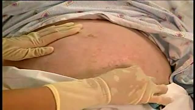 Fetal Heart Sounds78,951 views
 IVUS Vascular Imaging10,198 views
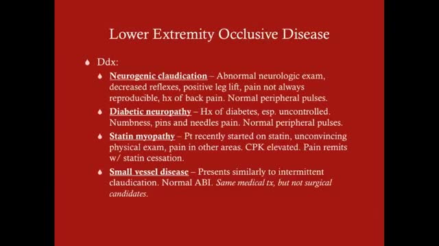 Lower Extremity Occlusive Disease1,448 views
Guide
What it is
A Doppler study uses the same probe and gel as a normal ultrasound scan, with the machine's Doppler mode switched on. You may hear a rhythmic whooshing from the speakers — that is your blood flow converted to sound — and see colour on the screen, conventionally red and blue for flow towards and away from the probe. The sonographer measures the speed of flow at chosen points and watches how the flow pattern changes. The test is painless, uses no radiation, and typically takes around half an hour depending on the area.
What it shows
Doppler studies answer flow questions that ordinary ultrasound cannot. In the legs, compression ultrasound with Doppler is the standard test for deep vein thrombosis (DVT) — a blood clot in the deep veins. In the neck, carotid Doppler measures narrowing of the arteries supplying the brain after a stroke or mini-stroke. Doppler also checks blood flow in the liver and kidney vessels, in transplanted organs, in the umbilical cord and placenta during pregnancy, and across heart valves as part of an echocardiogram. It cannot map vessels in the fine detail needed to plan most operations — CT or MR angiography does that.
How to prepare
Most Doppler studies need no preparation. For leg vein scans you will undress the leg or wear shorts; for neck (carotid) scans, wear a top that leaves the neck accessible and remove necklaces. Abdominal vessel studies may ask for fasting, because bowel gas blocks sound. The sonographer places gel on the skin and moves the probe along the vessel, sometimes pressing firmly — for leg veins, pressing to check the vein squashes flat is the key part of the test. You may be asked to tense your calf or bear down briefly to change the flow.
What the report words mean
Doppler reports talk about flow. 'Patent' means the vessel is open. 'Stenosis' is a narrowing, often given as a percentage for the carotid arteries because treatment decisions depend on it. 'Occlusion' means completely blocked. 'Reflux' in leg veins means blood flows backwards through faulty valves — the mechanism behind many varicose veins. 'Monophasic', 'biphasic', and 'triphasic' describe the shape of the arterial flow waveform; a healthy leg artery is usually triphasic, and losing phases suggests narrowing upstream. 'No DVT seen' in a leg report means the scanned veins compressed normally.
Common questions
How does the leg scan find a blood clot (DVT)?
A DVT scan is a compression ultrasound: the sonographer presses the probe on the leg veins at intervals. A healthy vein squashes flat completely; a vein containing clot does not compress fully. Doppler signals are used as well to check flow. It is the standard first test when a leg clot is suspected and involves no radiation.
Why does the report give a percentage for carotid narrowing?
The report gives a percentage narrowing because the degree of carotid narrowing is one of the main factors in deciding between medicines alone and a procedure to open or clear the artery. The percentage is an estimate from the speed of blood flow through the narrowed segment, not a direct measurement.
Is Doppler used in pregnancy?
Yes, during pregnancy a small handheld Doppler is routinely used to listen to the baby's heartbeat at antenatal appointments, and ultrasound scans use Doppler to check blood flow in the umbilical cord and the baby's heart when needed. These uses are considered safe and are a standard part of pregnancy care.
What happens if the Doppler finds a blockage?
A Doppler study measures flow, not anatomy in fine detail. If it suggests a significant narrowing or blockage, the next step is often a CT or MR angiogram to map the vessel precisely, especially if a procedure is being planned. Your clinician will explain which result would change your treatment.
More in Ultrasound
Related
Sources
- Vascular Ultrasound (RadiologyInfo.org (RSNA/ACR))
- Ultrasound — Doppler (MedlinePlus Medical Encyclopedia)
- Ultrasound scan (NHS)
This guide is general information, not medical advice, and does not replace advice from your own clinician. How a scan is done, and whether contrast is used, depends on your own medical history — always follow the instructions of the team performing your scan, and tell them about kidney problems, allergies, implants, and the medicines you take. A suspected blood clot with a painful, swollen leg needs same-day medical assessment — do not wait for a routine scan appointment.