
- Physical Examination
- Surgical Examination
- Ophthalmology
- Clinical Skills
- Orthopedics
- Surgery Videos
- Laparoscopy
- Pediatrics
- Funny Videos
- Cardiothoracic Surgery
- Nursing Videos
- Plastic Surgery
- Otorhinolaryngology
- Histology and Histopathology
- Neurosurgery
- Dermatology
- Pediatric Surgery
- Urology
- Dentistry
- Oncology and Cancers
- Anatomy Videos
- Health and Fitness
- Radiology
- Anaesthesia
- Physical Therapy
- Pharmacology
- Interventional Radiology
- Cardiology
- Endocrinology
- Gynecology
- Emergency Medicine
- Psychiatry and Psychology
- Childbirth Videos
- General Medical Videos
- Nephrology
- Physiology
- Diet and Food Health
- Diabetes Mellitus
- Neurology
- Women Health
- Osteoporosis
- Gastroenterology
- Pulmonology
- Hematology
- Rheumatology
- Toxicology
- Nuclear Medicine
- Infectious Diseases
- Vascular Disease
- Reproductive Health
- Burns and Wound Healing
- Kidney Zone Podcast
- Other
Intravenous urogram (IVU/IVP)
Category: Imaging & Scans · All imaging & scans
What it is
An intravenous urogram is an X-ray examination of the kidneys, ureters and bladder using contrast dye injected into a vein, which is filtered by the kidneys so the urinary tract shows up on a timed series of pictures. In many centres CT urography has replaced it, but it is still used in some settings.
Why it is done
It may be used to investigate blood in the urine, kidney stones, repeated urinary infections or blockages of the urinary tract.
How to prepare
You may be asked not to eat for a few hours and sometimes to take a mild laxative beforehand so bowel gas does not hide the pictures. Tell the team about allergies, asthma, diabetes medicines and kidney problems.
What happens during the procedure
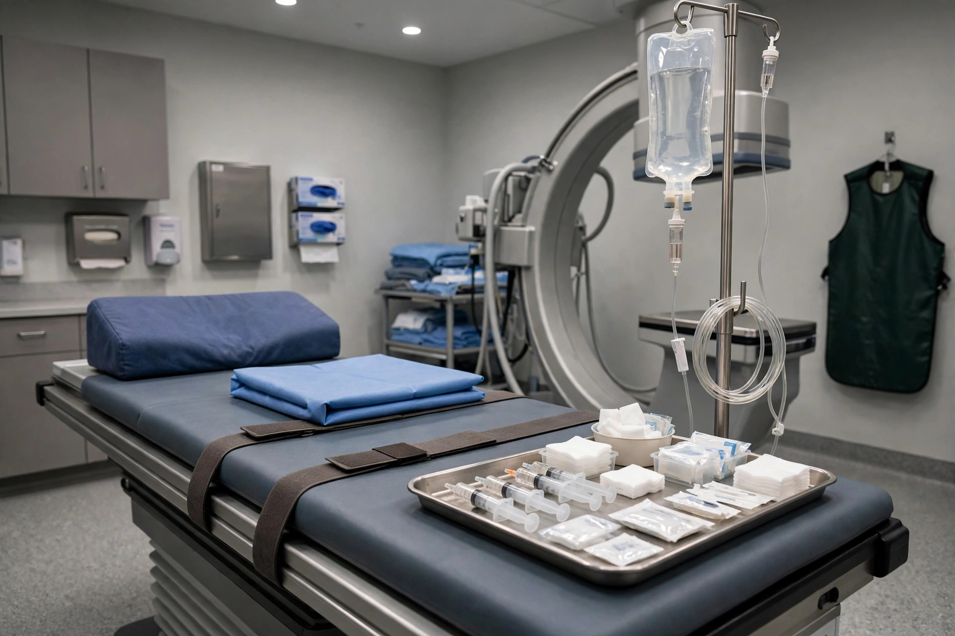
A dye injection is given into a vein in the arm, then a series of X-rays is taken over 20 to 60 minutes as the dye passes through the kidneys and down to the bladder. You may be asked to empty your bladder partway through for final pictures.
Afterwards — recovery & results
You can usually go home afterwards and eat and drink normally. Drinking water helps flush out the dye. The report goes to your doctor.
Risks & considerations
Risks include an allergic-type reaction to the dye and effects on the kidneys, so kidney function is checked first. The radiation dose is modest. If your centre offers CT urography instead, it gives more detail at a higher dose.
Related guides on MedicalVideos.com
Frequently asked questions
Why several X-rays over an hour?
The pictures are timed to catch the dye at each stage — filling the kidneys, travelling down the ureters, and collecting in the bladder — so the whole tract is assessed.
I have a kidney problem — can I still have it?
The team will check your kidney function first and may choose a different test if the dye is not suitable for you.
Related procedures — Imaging & Scans
Browse the I section of the A–Z list or return to the Procedures & Tests hub.