
- Physical Examination
- Surgical Examination
- Ophthalmology
- Clinical Skills
- Orthopedics
- Surgery Videos
- Laparoscopy
- Pediatrics
- Funny Videos
- Cardiothoracic Surgery
- Nursing Videos
- Plastic Surgery
- Otorhinolaryngology
- Histology and Histopathology
- Neurosurgery
- Dermatology
- Pediatric Surgery
- Urology
- Dentistry
- Oncology and Cancers
- Anatomy Videos
- Health and Fitness
- Radiology
- Anaesthesia
- Physical Therapy
- Pharmacology
- Interventional Radiology
- Cardiology
- Endocrinology
- Gynecology
- Emergency Medicine
- Psychiatry and Psychology
- Childbirth Videos
- General Medical Videos
- Nephrology
- Physiology
- Diet and Food Health
- Diabetes Mellitus
- Neurology
- Women Health
- Osteoporosis
- Gastroenterology
- Pulmonology
- Hematology
- Rheumatology
- Toxicology
- Nuclear Medicine
- Infectious Diseases
- Vascular Disease
- Reproductive Health
- Burns and Wound Healing
- Kidney Zone Podcast
- Other
Carotid Doppler ultrasound
Category: Imaging & Scans · All imaging & scans
What it is
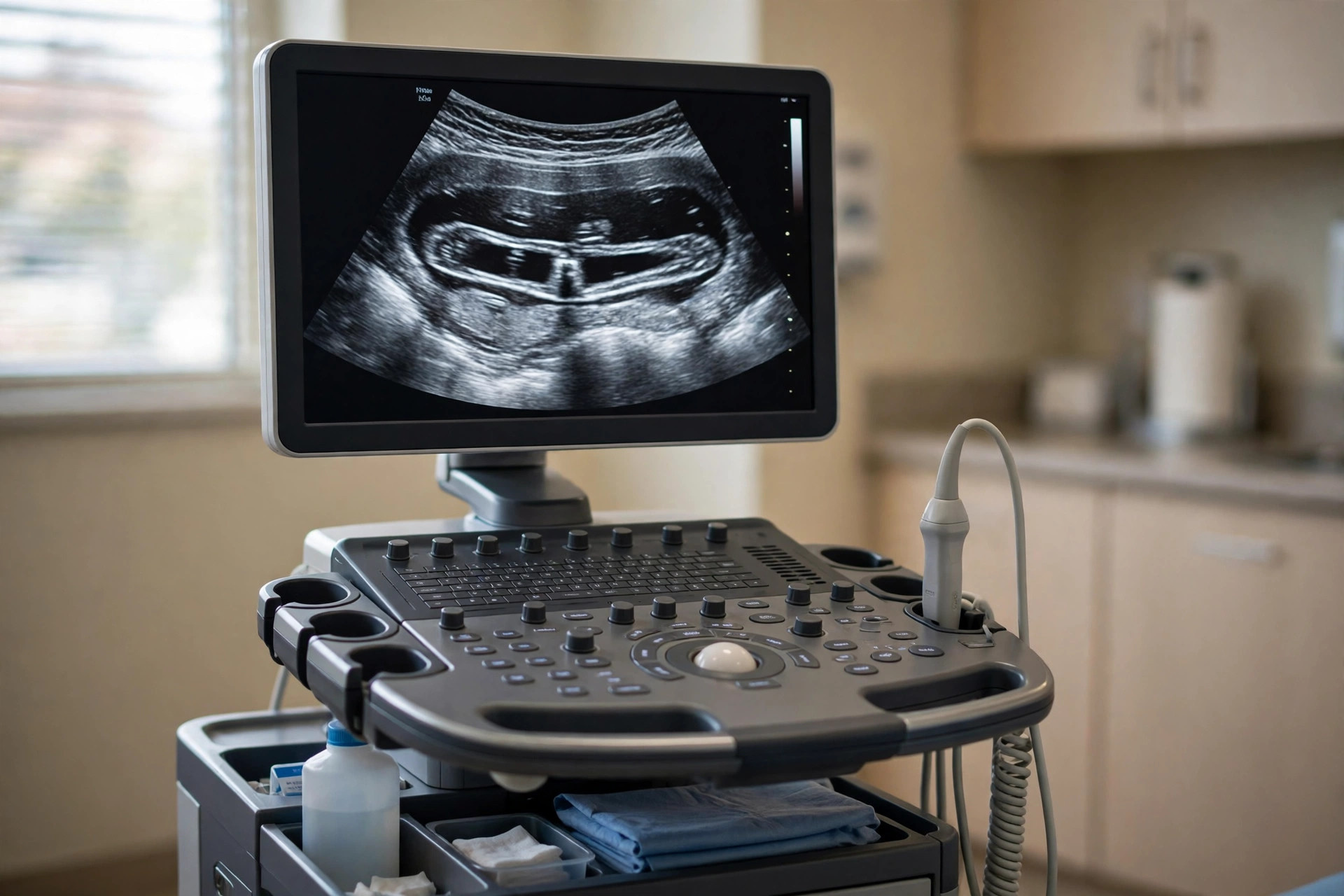
A carotid Doppler ultrasound uses sound waves to picture the carotid arteries in the neck and measure how blood flows through them, looking for narrowing caused by fatty deposits.
Why it is done
It is often arranged after a stroke or mini-stroke (TIA), or when a narrowing sound (bruit) is heard in the neck, to help decide whether treatment of the artery is advised.
How to prepare
There is no special preparation and you can eat, drink and take your medicines as usual.
What happens during the procedure
You lie on a couch with your neck slightly extended. Gel is applied and the probe is moved along each side of the neck while the machine measures flow; you may hear a pulsing sound from the machine's speaker. It takes about 20 to 30 minutes and is painless.
Afterwards — recovery & results
You can go home straight away. Results are usually sent to your doctor or the stroke team promptly, because they can influence decisions about treatment.
Risks & considerations
There are no known risks from the ultrasound itself. The main limitation is that it estimates narrowing rather than picturing it directly, so a CT or MR angiogram is sometimes advised to confirm the degree of narrowing.
Related guides on MedicalVideos.com
Frequently asked questions
What does the whooshing sound mean?
The machine converts blood flow into sound. Changes in pitch help the operator judge how fast blood is moving, which relates to how narrow the artery is.
If narrowing is found, will I need an operation?
Not necessarily. Many narrowings are treated with medicines and risk-factor control. Your specialist will discuss the options based on the degree of narrowing and your symptoms.
Related procedures — Imaging & Scans
Browse the C section of the A–Z list or return to the Procedures & Tests hub.