
- Physical Examination
- Surgical Examination
- Ophthalmology
- Clinical Skills
- Orthopedics
- Surgery Videos
- Laparoscopy
- Pediatrics
- Funny Videos
- Cardiothoracic Surgery
- Nursing Videos
- Plastic Surgery
- Otorhinolaryngology
- Histology and Histopathology
- Neurosurgery
- Dermatology
- Pediatric Surgery
- Urology
- Dentistry
- Oncology and Cancers
- Anatomy Videos
- Health and Fitness
- Radiology
- Anaesthesia
- Physical Therapy
- Pharmacology
- Interventional Radiology
- Cardiology
- Endocrinology
- Gynecology
- Emergency Medicine
- Psychiatry and Psychology
- Childbirth Videos
- General Medical Videos
- Nephrology
- Physiology
- Diet and Food Health
- Diabetes Mellitus
- Neurology
- Women Health
- Osteoporosis
- Gastroenterology
- Pulmonology
- Hematology
- Rheumatology
- Toxicology
- Nuclear Medicine
- Infectious Diseases
- Vascular Disease
- Reproductive Health
- Burns and Wound Healing
- Kidney Zone Podcast
- Other
Imaging & Scans
Scans and imaging tests: X-rays, CT, MRI, ultrasound in its many forms, mammography, bone-density scanning and nuclear medicine. These pages explain what each scan shows, how to prepare, what happens in the scanner room, and what the pictures can and cannot tell your team.
This page lists all 30 of them in the guide. Open any procedure for preparation, step-by-step what happens, recovery advice and frequently asked questions.
 Abdominal ultrasoundImaging & Scans
 Angiography (catheter angiogram)Imaging & Scans
 Barium swallow and mealImaging & Scans
 Breast ultrasoundImaging & Scans
 Carotid Doppler ultrasoundImaging & Scans
 Chest X-rayImaging & Scans
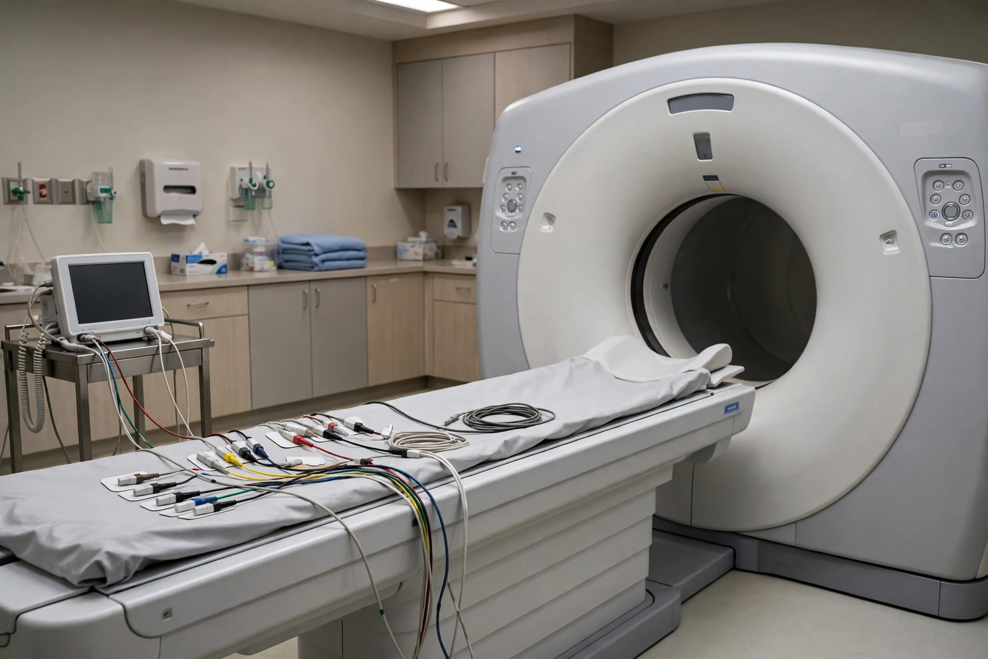 Coronary CT angiographyImaging & Scans
 CT head scanImaging & Scans
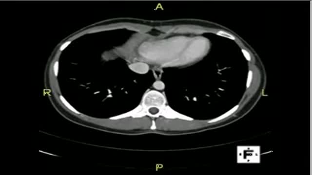 CT scan — abdomen and pelvisImaging & Scans
 CT scan — chestImaging & ScansCT scan — headImaging & Scans
 DEXA bone density scanImaging & Scans
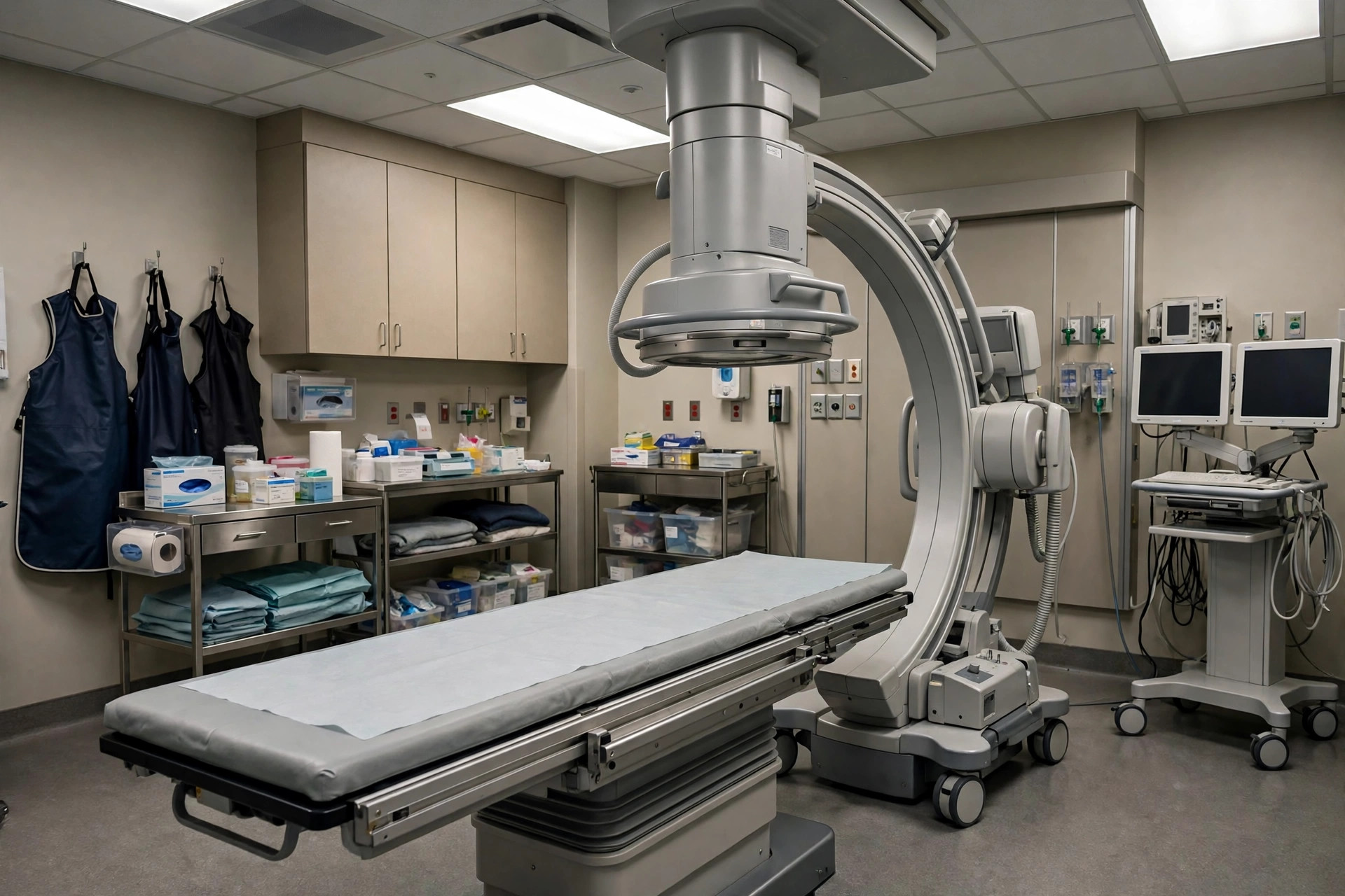 Fluoroscopy (real-time X-ray)Imaging & Scans
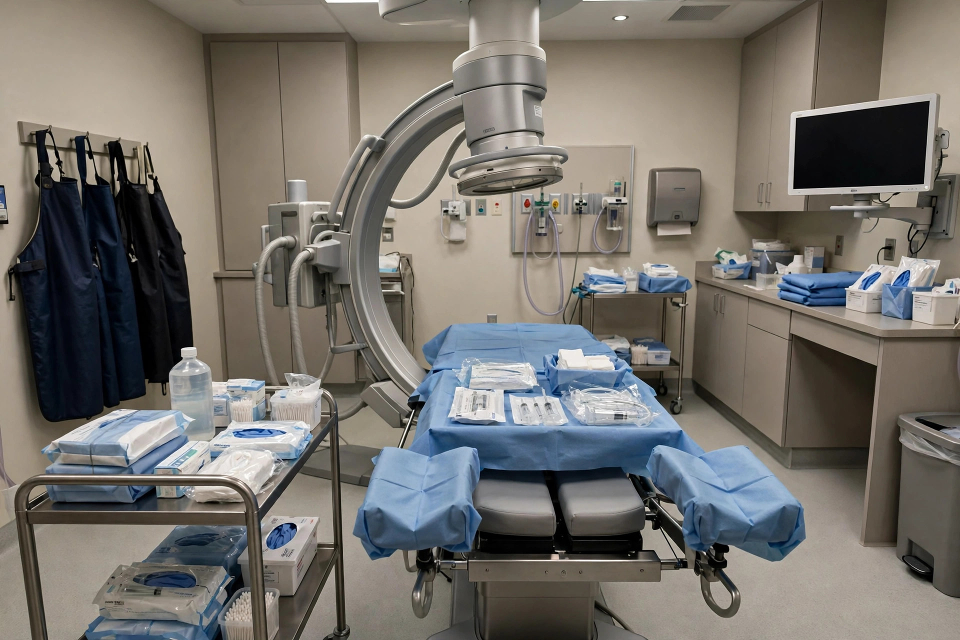 Hysterosalpingogram (HSG)Imaging & Scans
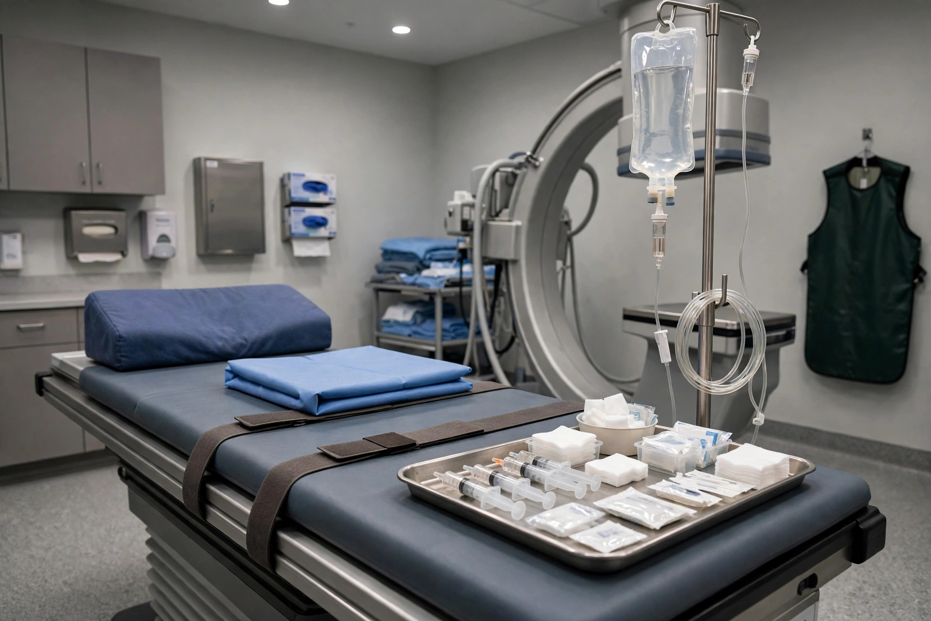 Intravenous urogram (IVU/IVP)Imaging & Scans
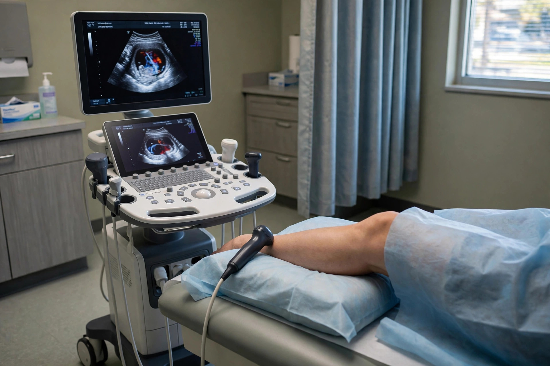 Leg Doppler ultrasound (vein scan)Imaging & Scans
 MammographyImaging & Scans
 MRI brain scanImaging & ScansMRI scan — brainImaging & Scans
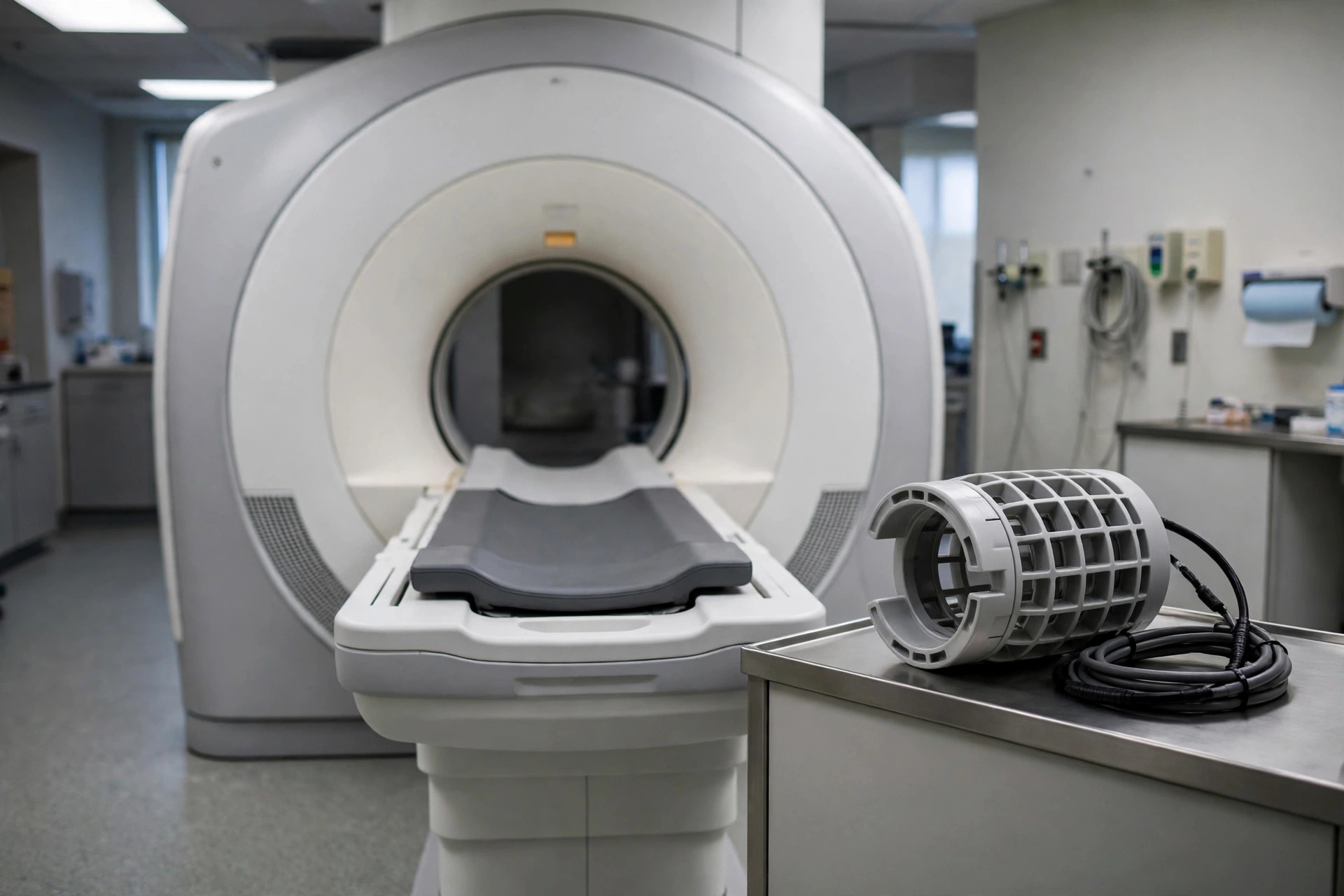 MRI scan — kneeImaging & Scans
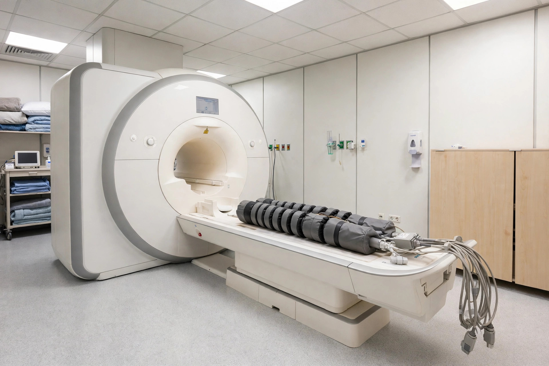 MRI scan — spineImaging & Scans
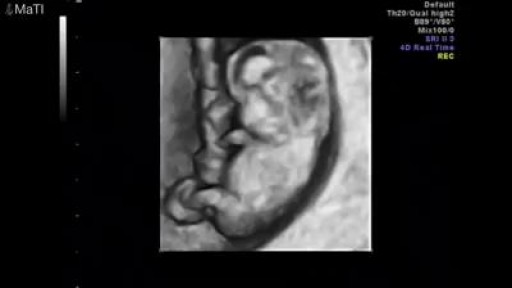 Obstetric ultrasound (pregnancy scan)Imaging & Scans
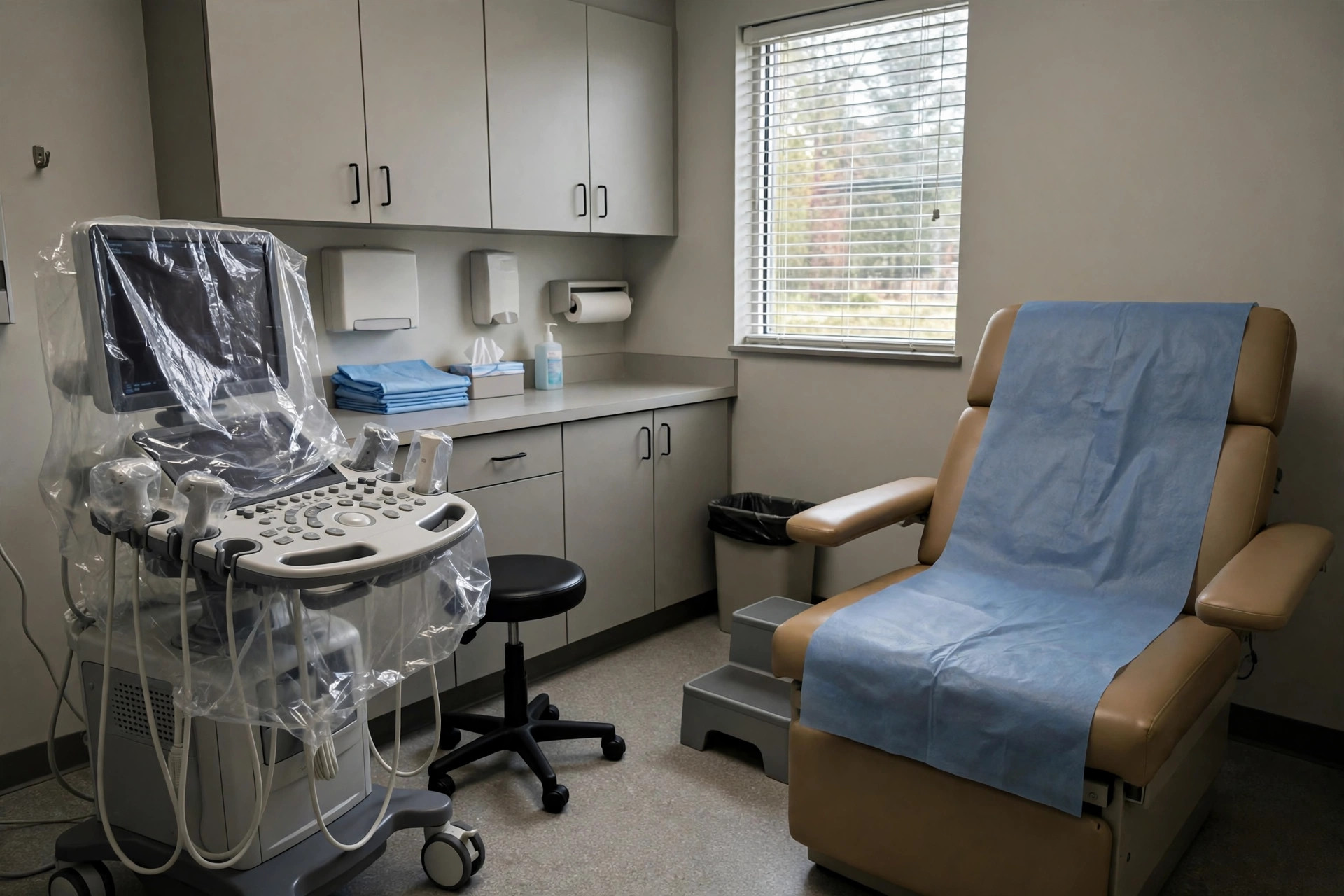 Pelvic ultrasoundImaging & Scans
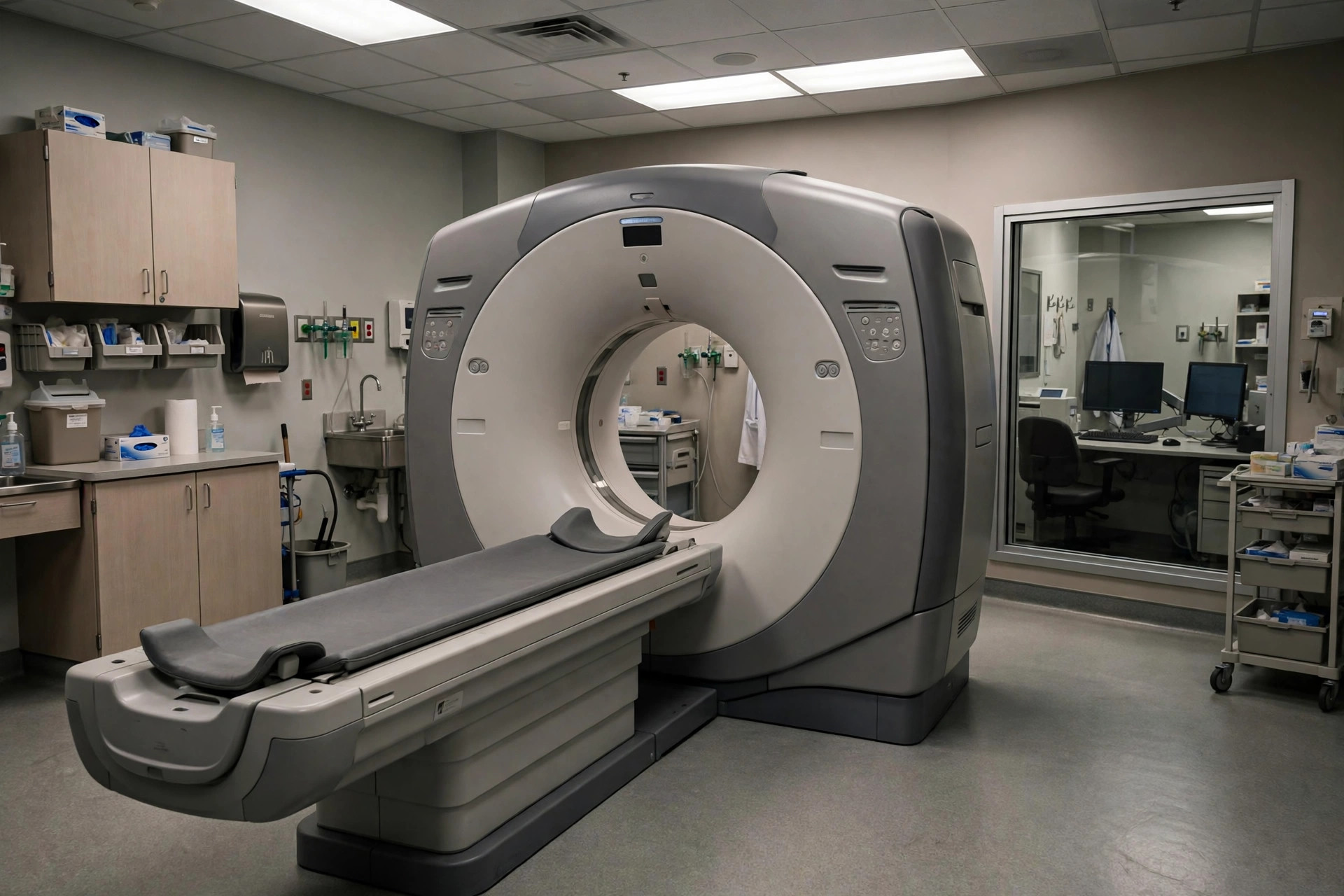 PET scanImaging & Scans
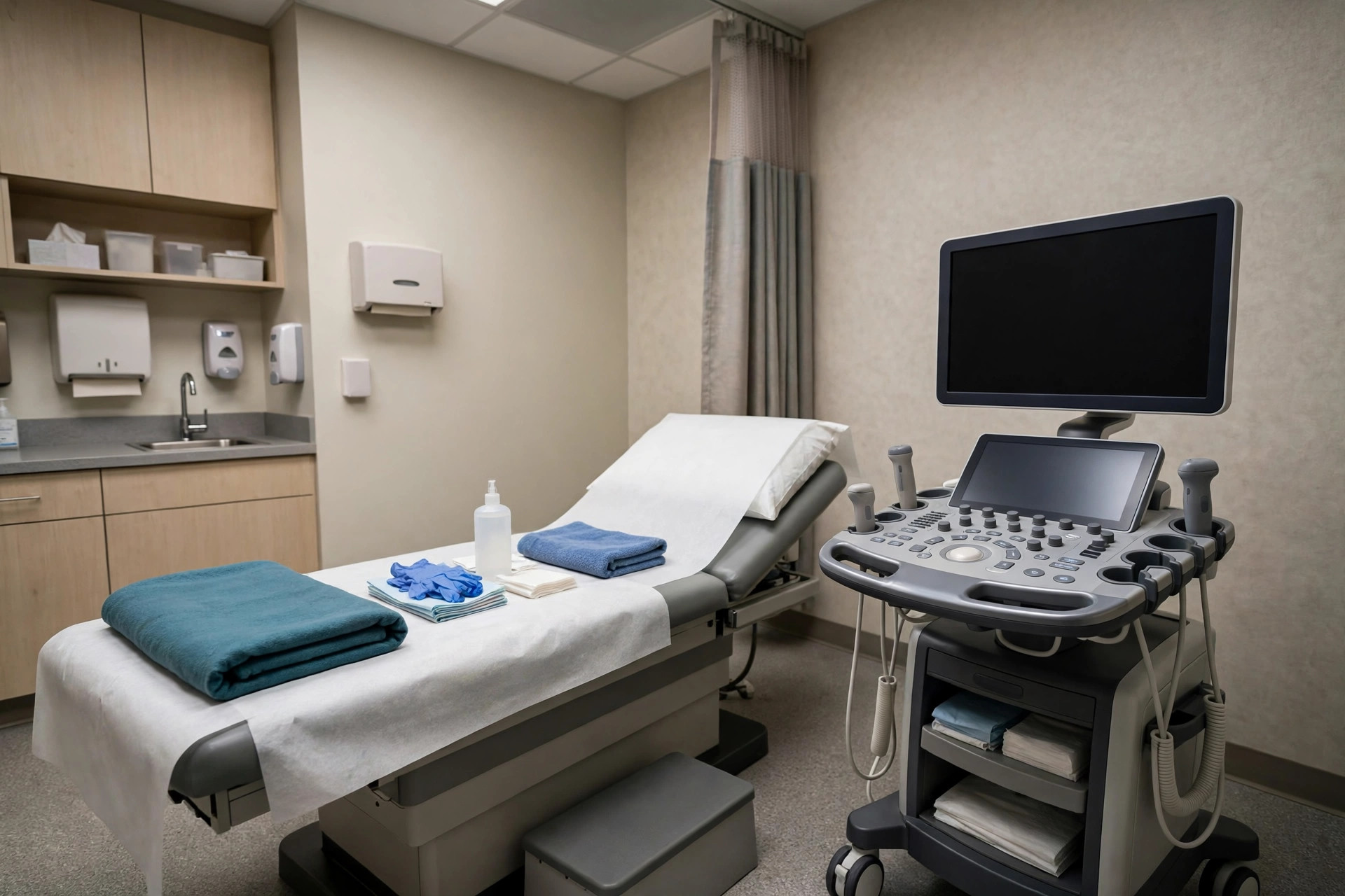 Scrotal ultrasoundImaging & Scans
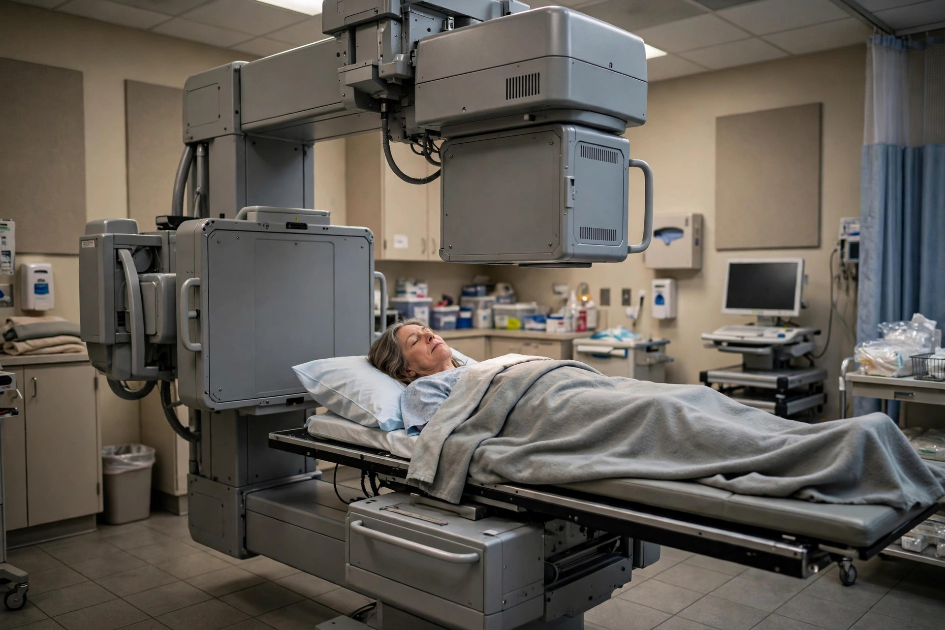 SPECT and nuclear medicine scansImaging & Scans
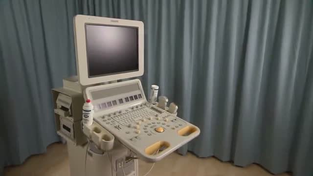 Thyroid ultrasoundImaging & Scans
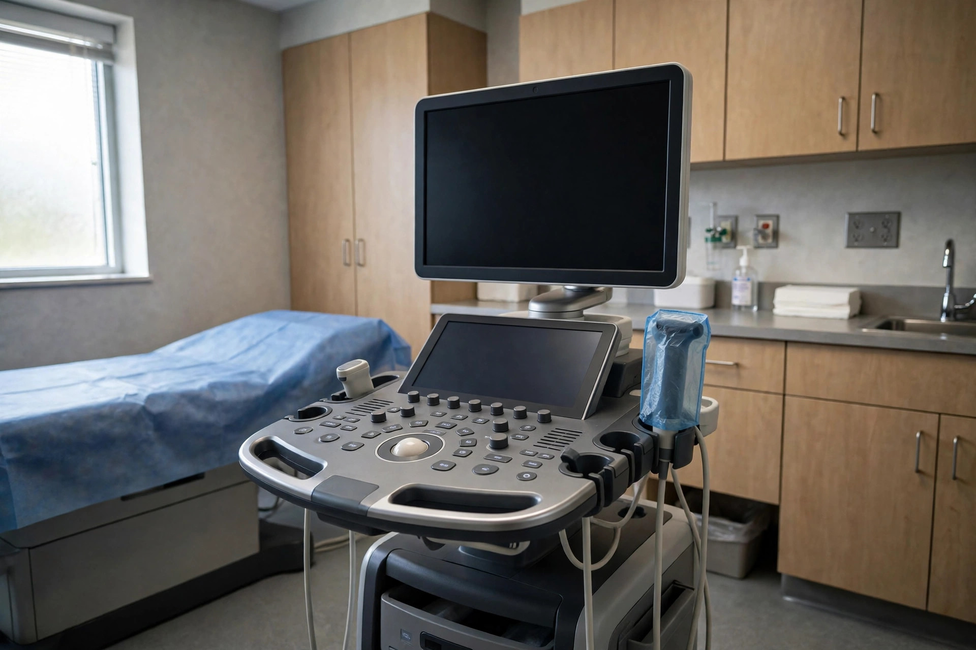 Transrectal ultrasound and prostate biopsy guidanceImaging & Scans
 Voiding cystourethrogram (MCUG / VCUG)Imaging & Scans
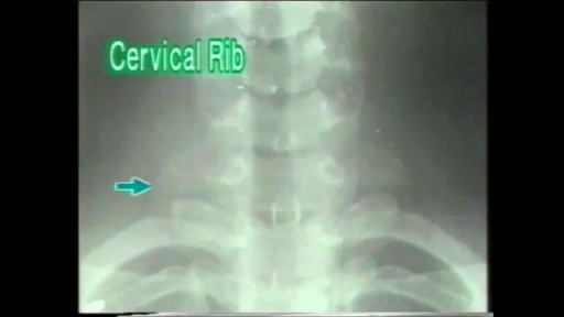 X-ray (general)Imaging & Scans
| Procedure / test | Category | What it is |
|---|---|---|
| Abdominal ultrasound | Imaging & Scans | An abdominal ultrasound uses high-frequency sound waves from a hand-held probe to create moving pictures of the liver, gallbladder, kidneys, pancreas, spleen and major blood vessels. It uses no radiation. |
| Angiography (catheter angiogram) | Imaging & Scans | An angiogram is an X-ray procedure in which contrast dye is injected through a fine tube (catheter) placed into a blood vessel, so that arteries or veins and any narrowings or blockages show up clearly. |
| Barium swallow and meal | Imaging & Scans | A barium swallow or barium meal is an X-ray examination in which you drink a white, chalky liquid containing barium so the gullet, stomach and upper bowel show up clearly on moving X-ray pictures (fluoroscopy). |
| Breast ultrasound | Imaging & Scans | A breast ultrasound uses sound waves to picture breast tissue. It is particularly good at telling whether a lump is fluid-filled (a cyst) or solid, and is often the first imaging test for breast symptoms in younger women. |
| Carotid Doppler ultrasound | Imaging & Scans | A carotid Doppler ultrasound uses sound waves to picture the carotid arteries in the neck and measure how blood flows through them, looking for narrowing caused by fatty deposits. |
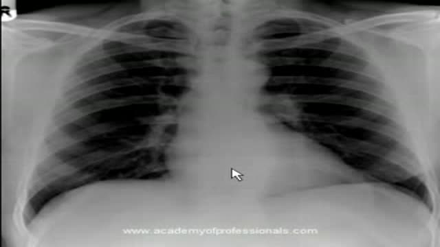
| Chest X-ray | Imaging & Scans | A chest X-ray is a quick picture of the chest made with a small amount of radiation. It shows the lungs, the heart outline, the ribs and the upper spine on one or two images. |
| Coronary CT angiography | Imaging & Scans | Coronary CT angiography is a CT scan that uses contrast dye to picture the coronary arteries — the vessels that supply the heart muscle — and look for narrowing caused by fatty deposits. |
| CT head scan | Imaging & Scans | A CT head scan takes X-ray pictures in slices through the brain, giving fast, clear images of bleeding, skull fractures, tumours and major strokes. It is the first-line emergency brain scan because it is quick and widely available. |
| CT scan — abdomen and pelvis | Imaging & Scans | A CT scan of the abdomen and pelvis creates detailed cross-section images of the bowel, liver, kidneys, pancreas, bladder and other organs in the belly and pelvis. |
| CT scan — chest | Imaging & Scans | A CT scan of the chest builds detailed cross-section pictures of the lungs, airways, heart and the large blood vessels in the chest, using X-rays and a computer. |
| CT scan — head | Imaging & Scans | A CT (computed tomography) scan of the head uses X-rays taken from many angles and a computer to build detailed cross-section pictures of the brain, skull and sinuses. |
| DEXA bone density scan | Imaging & Scans | A DEXA (dual-energy X-ray absorptiometry) scan measures bone density using very low-dose X-rays, usually at the hip and spine. The result helps assess bone strength and the risk of fractures. |
| Fluoroscopy (real-time X-ray) | Imaging & Scans | Fluoroscopy is a moving X-ray picture — like an X-ray video — that lets doctors watch structures and instruments in real time. It is used to guide many procedures and to watch swallowing, bowel movement and joint movement as they happen. |
| Hysterosalpingogram (HSG) | Imaging & Scans | A hysterosalpingogram (HSG) is an X-ray test in which a small amount of dye is passed through the neck of the womb so the womb cavity and fallopian tubes can be seen and checked for blockages. |
| Intravenous urogram (IVU/IVP) | Imaging & Scans | An intravenous urogram is an X-ray examination of the kidneys, ureters and bladder using contrast dye injected into a vein, which is filtered by the kidneys so the urinary tract shows up on a timed series of pictures. In many centres CT urography has replaced it, but it is still used in some settings. |
| Leg Doppler ultrasound (vein scan) | Imaging & Scans | A leg Doppler (venous duplex) ultrasound uses sound waves to check the deep veins of the leg for a blood clot (deep vein thrombosis, DVT) and to assess vein valves in varicose vein problems. |
| Mammography | Imaging & Scans | A mammogram is a specialised low-dose X-ray of the breast. Each breast is briefly compressed between two flat plates so the tissue spreads out and detailed pictures can be taken. |
| MRI brain scan | Imaging & Scans | An MRI of the brain uses strong magnets and radio waves to produce detailed pictures of the brain and its structures without X-rays. Sequences can highlight tumours, stroke, inflammation, infection, bleeding products and structural abnormalities, and contrast dye may be given to show blood vessels and areas of inflammation or tumour activity. |
| MRI scan — brain | Imaging & Scans | An MRI (magnetic resonance imaging) scan of the brain uses a strong magnet and radio waves — not X-rays — to build very detailed pictures of the brain and its blood vessels. |
| MRI scan — knee | Imaging & Scans | An MRI scan of the knee uses a magnet and radio waves to produce detailed pictures of the cartilage, ligaments, tendons and bones inside the knee joint. |
| MRI scan — spine | Imaging & Scans | An MRI scan of the spine uses a magnet and radio waves to picture the spinal bones, discs, spinal cord and nerves in fine detail, without using X-rays. |
| Obstetric ultrasound (pregnancy scan) | Imaging & Scans | An obstetric ultrasound uses sound waves to picture the baby in the womb. Routine scans are usually offered in early pregnancy (the dating scan) and again around the middle of pregnancy (the anomaly scan), with extra scans if needed. |
| Pelvic ultrasound | Imaging & Scans | A pelvic ultrasound uses sound waves to picture the womb, ovaries and surrounding structures. It may be done through the tummy (transabdominal), through the vagina (transvaginal), or both, depending on what needs to be seen. |
| PET scan | Imaging & Scans | A PET (positron emission tomography) scan uses a small amount of a radioactive tracer, usually a form of sugar, to show how active tissues are. It is usually combined with a CT scan in the same machine (PET-CT). |
| Scrotal ultrasound | Imaging & Scans | A scrotal ultrasound uses sound waves to picture the testicles and surrounding structures, including blood flow. It is a quick, painless way to assess lumps and pain. |
| SPECT and nuclear medicine scans | Imaging & Scans | Nuclear medicine scans inject a tiny amount of radioactive tracer and use a gamma camera to picture how organs are working rather than just how they look — for example bone scans (tracer gathers where bone is active), thyroid scans, kidney (renogram) scans and lung perfusion scans. SPECT adds 3D (tomographic) imaging. |
| Thyroid ultrasound | Imaging & Scans | A thyroid ultrasound uses sound waves to picture the thyroid gland in the front of the neck, showing its size and any lumps (nodules) within it. |
| Transrectal ultrasound and prostate biopsy guidance | Imaging & Scans | A transrectal ultrasound (TRUS) uses a slim ultrasound probe placed in the back passage to picture the prostate gland in detail. It is often combined with taking small tissue samples (biopsies) from the prostate. |
| Voiding cystourethrogram (MCUG / VCUG) | Imaging & Scans | A voiding cystourethrogram is an X-ray study of the bladder and urethra: contrast dye is passed into the bladder through a small catheter, and X-ray pictures are taken while the bladder fills and while it empties (voiding). It shows whether urine flows backwards towards the kidneys (vesicoureteric reflux) and whether the outflow is normal. |
| X-ray (general) | Imaging & Scans | An X-ray is a picture of the inside of the body made with a small, focused amount of radiation. Bones show up white and are seen very clearly, which makes X-rays the usual first test for suspected broken bones and joint problems. |
Please note: This page is educational information about a medical procedure or test. It does not replace the instructions of your own care team, who know your situation and whose preparation, fasting and medication advice you should follow if it differs from anything here. If you have symptoms that worry you, or an appointment you do not understand, contact your clinical team.