
- Physical Examination
- Surgical Examination
- Ophthalmology
- Clinical Skills
- Orthopedics
- Surgery Videos
- Laparoscopy
- Pediatrics
- Funny Videos
- Cardiothoracic Surgery
- Nursing Videos
- Plastic Surgery
- Otorhinolaryngology
- Histology and Histopathology
- Neurosurgery
- Dermatology
- Pediatric Surgery
- Urology
- Dentistry
- Oncology and Cancers
- Anatomy Videos
- Health and Fitness
- Radiology
- Anaesthesia
- Physical Therapy
- Pharmacology
- Interventional Radiology
- Cardiology
- Endocrinology
- Gynecology
- Emergency Medicine
- Psychiatry and Psychology
- Childbirth Videos
- General Medical Videos
- Nephrology
- Physiology
- Diet and Food Health
- Diabetes Mellitus
- Neurology
- Women Health
- Osteoporosis
- Gastroenterology
- Pulmonology
- Hematology
- Rheumatology
- Toxicology
- Nuclear Medicine
- Infectious Diseases
- Vascular Disease
- Reproductive Health
- Burns and Wound Healing
- Kidney Zone Podcast
- Other
OCT scan (optical coherence tomography)
Category: Eye Procedures & Tests · All eye procedures & tests
What it is
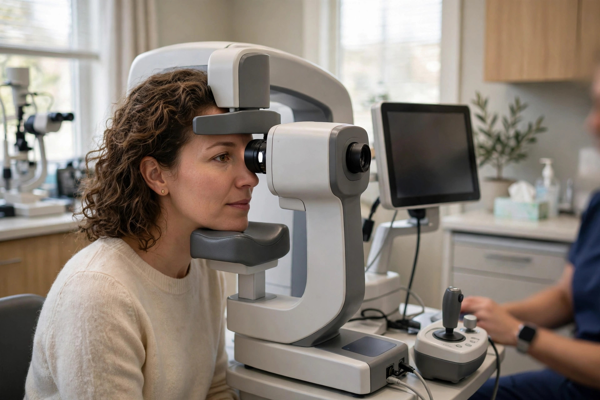
OCT is a quick, non-contact scan that uses light waves to build a cross-section picture of the retina (and can also image the cornea and optic nerve), showing its layers in microscopic detail.
Why it is done
It is central to diagnosing and monitoring macular degeneration, diabetic macular oedema, macular holes and membranes, glaucoma (nerve fibre measurements) and many other retinal conditions, and to guiding injection and laser treatment decisions.
How to prepare
No preparation is needed; sometimes dilating drops are used, which blur vision for a few hours. Bring your glasses and a list of eye medicines.
What happens during the procedure
You sit with your chin on a rest and look at a target while the machine scans each eye — no contact with the eye, no discomfort. It takes a few minutes per eye.
Afterwards — recovery & results
You can return to normal activities immediately (once any dilation wears off for driving). Results are usually reviewed in the same clinic visit and compared with your previous scans to detect change.
Risks & considerations
There are no known risks from OCT — it uses low-power light. Its limitation is that it is one test among several; findings are interpreted with examination and vision testing.
Related guides on MedicalVideos.com
Frequently asked questions
Is OCT the same as the scan at the optician?
Often yes — many opticians offer OCT. Hospital scans use similar technology, interpreted alongside your full eye assessment.
Why repeat the scan every visit?
Comparing layers over time shows whether fluid or thinning is improving, stable or worsening, which is how treatment intervals are decided.
Related procedures — Eye Procedures & Tests
Browse the O section of the A–Z list or return to the Procedures & Tests hub.