
- Physical Examination
- Surgical Examination
- Ophthalmology
- Clinical Skills
- Orthopedics
- Surgery Videos
- Laparoscopy
- Pediatrics
- Funny Videos
- Cardiothoracic Surgery
- Nursing Videos
- Plastic Surgery
- Otorhinolaryngology
- Histology and Histopathology
- Neurosurgery
- Dermatology
- Pediatric Surgery
- Urology
- Dentistry
- Oncology and Cancers
- Anatomy Videos
- Health and Fitness
- Radiology
- Anaesthesia
- Physical Therapy
- Pharmacology
- Interventional Radiology
- Cardiology
- Endocrinology
- Gynecology
- Emergency Medicine
- Psychiatry and Psychology
- Childbirth Videos
- General Medical Videos
- Nephrology
- Physiology
- Diet and Food Health
- Diabetes Mellitus
- Neurology
- Women Health
- Osteoporosis
- Gastroenterology
- Pulmonology
- Hematology
- Rheumatology
- Toxicology
- Nuclear Medicine
- Infectious Diseases
- Vascular Disease
- Reproductive Health
- Burns and Wound Healing
- Kidney Zone Podcast
- Other
Transcranial Doppler
Category: Brain, Nerve & Sleep Tests · All brain, nerve & sleep tests
What it is
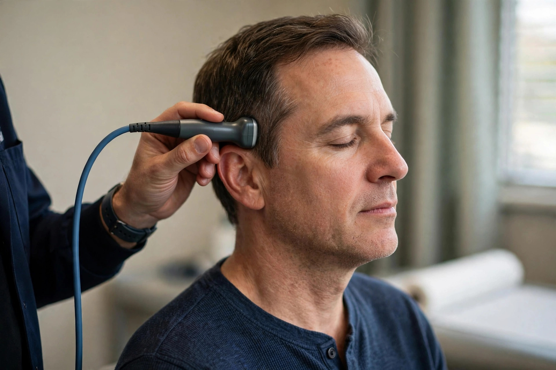
Transcranial Doppler uses ultrasound through the thin bone of the temple to measure blood flow speeds in the brain's major arteries in real time. It can detect narrowing, monitor spasm after brain haemorrhage, find right-to-left shunts (with a bubble test using agitated saline) and monitor for tiny emboli.
Why it is done
Uses include monitoring after subarachnoid haemorrhage, sickle cell disease stroke-risk screening in children, stroke work-up (bubble study for patent foramen ovale), and intra-operative monitoring.
How to prepare
No preparation is needed. You lie or sit comfortably; the test cannot be done well in a small percentage of people whose skull window blocks ultrasound — alternative tests are used then.
What happens during the procedure
A probe with gel is held at the temple (and sometimes under the skull base or over the eye socket with very low power settings) while flow signals are identified by sound and waveform. For a bubble study, agitated saline is injected into an arm vein while you perform a straining manoeuvre, and the machine listens for bubbles crossing to the brain arteries. The test takes about 30 to 45 minutes.
Afterwards — recovery & results
You can leave immediately. Results are reported to your team; a positive bubble study feeds into stroke-cause discussions (for example whether a heart hole needs closure).
Risks & considerations
Ultrasound is harmless. The bubble study's saline bubbles are tiny and dissolve; serious reactions are essentially unheard of in routine practice.
Related guides on MedicalVideos.com
Frequently asked questions
What does a 'positive bubble study' mean?
Bubbles that should be filtered by the lungs reached the brain arteries, implying a right-to-left shunt — usually a patent foramen ovale, a common finding whose significance depends on your stroke story.
Why monitor after a brain haemorrhage daily?
Arteries can go into spasm days after a subarachnoid bleed, starving the brain. Rising flow velocities warn the team to intervene before damage occurs.
Related procedures — Brain, Nerve & Sleep Tests
Browse the T section of the A–Z list or return to the Procedures & Tests hub.