
- Physical Examination
- Surgical Examination
- Ophthalmology
- Clinical Skills
- Orthopedics
- Surgery Videos
- Laparoscopy
- Pediatrics
- Funny Videos
- Cardiothoracic Surgery
- Nursing Videos
- Plastic Surgery
- Otorhinolaryngology
- Histology and Histopathology
- Neurosurgery
- Dermatology
- Pediatric Surgery
- Urology
- Dentistry
- Oncology and Cancers
- Anatomy Videos
- Health and Fitness
- Radiology
- Anaesthesia
- Physical Therapy
- Pharmacology
- Interventional Radiology
- Cardiology
- Endocrinology
- Gynecology
- Emergency Medicine
- Psychiatry and Psychology
- Childbirth Videos
- General Medical Videos
- Nephrology
- Physiology
- Diet and Food Health
- Diabetes Mellitus
- Neurology
- Women Health
- Osteoporosis
- Gastroenterology
- Pulmonology
- Hematology
- Rheumatology
- Toxicology
- Nuclear Medicine
- Infectious Diseases
- Vascular Disease
- Reproductive Health
- Burns and Wound Healing
- Kidney Zone Podcast
- Other
Pterygium excision: What to Expect & Recovery
A pterygium is a fleshy growth from the white of the eye onto the cornea, driven by sun exposure. Excision removes it and usually covers the area with a tiny graft of your own conjunctiva, which greatly reduces recurrence. Day-case surgery under local anaesthetic; the eye is red and irritable for a few weeks.
Before surgery: how to prepare
- Tell the surgeon about blood thinners and eye allergies; bring sunglasses for the journey home — the eye will be light-sensitive.
- Arrange a lift home; the eye is usually padded initially.
- Recurrence is the central issue in this surgery: ask whether a conjunctival graft (with stitches or tissue glue) is planned, as grafting roughly quarters recurrence compared with bare excision in published series — your surgeon will describe their technique.
- If the pterygium is quiet and not threatening vision, surgery may be optional; if it is growing toward the pupil or distorting vision, removal is advised before it advances further.
The day of surgery
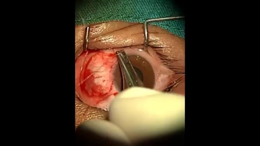
Under local anaesthetic, the growth is dissected off the cornea and the underlying sclera, the cornea is smoothed, and the bare area is covered with a thin graft of healthy conjunctiva from under the upper lid, fixed with fine stitches or fibrin glue. The procedure takes about 30 to 45 minutes, and you go home with a pad and drops.
In hospital
Recovery timeline
Every recovery is individual, but most follow this general arc:
Warning signs: when to get help
- Increasing pain, especially severe pain in the first days (the graft and cornea should be checked)
- Vision worsening
- Discharge with increasing redness after initial improvement (infection)
- The graft looking lifted, white or displaced at your check — report between visits if the eye suddenly worsens
- New fleshy growth at the site months later (recurrence)
Related topics
Watch: real operation videos
From the MedicalVideos library. Some surgical videos are graphic and intended for education.
Frequently asked questions
Why use a graft? Can it not just be scraped off?
Bare excision leaves raw sclera, and the body's healing response frequently regrows the pterygium — older series reported recurrence in a large minority. Covering the area with healthy conjunctival tissue gives the surface a normal template and cuts recurrence substantially, at the cost of a slightly longer operation.
Will my vision improve after removal?
If the pterygium was pulling on the cornea (causing astigmatism) or nearing the visual axis, removing it usually improves clarity over weeks as the cornea reshapes. Long-standing pterygia can leave a faint corneal scar that limits the final sharpness — your surgeon can usually predict this from the pre-operative examination.
Medically reviewed by Dr. Mohamed Ibrahim, Assistant Professor of Medicine, Transplant Nephrology, University of Maryland School of Medicine. Last reviewed: 12 November 2026.