- Physical Examination
- Surgical Examination
- Ophthalmology
- Clinical Skills
- Orthopedics
- Surgery Videos
- Laparoscopy
- Pediatrics
- Funny Videos
- Cardiothoracic Surgery
- Nursing Videos
- Plastic Surgery
- Otorhinolaryngology
- Histology and Histopathology
- Neurosurgery
- Dermatology
- Pediatric Surgery
- Urology
- Dentistry
- Oncology and Cancers
- Anatomy Videos
- Health and Fitness
- Radiology
- Anaesthesia
- Physical Therapy
- Pharmacology
- Interventional Radiology
- Cardiology
- Endocrinology
- Gynecology
- Emergency Medicine
- Psychiatry and Psychology
- Childbirth Videos
- General Medical Videos
- Nephrology
- Physiology
- Diet and Food Health
- Diabetes Mellitus
- Neurology
- Women Health
- Osteoporosis
- Gastroenterology
- Pulmonology
- Hematology
- Rheumatology
- Toxicology
- Nuclear Medicine
- Infectious Diseases
- Vascular Disease
- Reproductive Health
- Burns and Wound Healing
- Kidney Zone Podcast
- Other
Eye
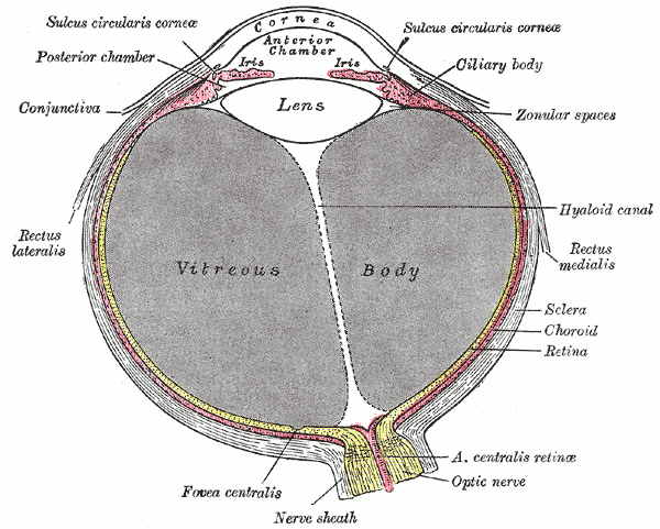
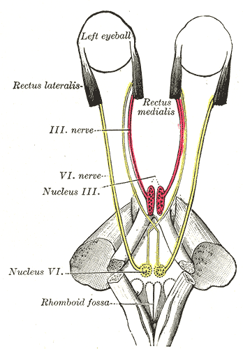
Gray's Anatomy (1918) plate, public domain
The eye is a fluid-filled sphere about 2.5 centimetres across that turns light into neural signals. The cornea and lens focus images onto the retina at the back; the iris controls the pupil's size to regulate light; and muscles move the eye in its socket. The front segment holds clear aqueous fluid, the back a gel (vitreous) that maintains the eye's shape.
Key facts
- The cornea provides most of the eye's focusing power; the lens fine-tunes it.
- The eye's lens changes shape to focus near objects (accommodation).
- The retina contains about 120 million rods and 6 million cones.
- Each eye has a blind spot where the optic nerve leaves.
- The eyeball's pressure is maintained by continuous production and drainage of aqueous fluid.
What can go wrong
Common eye problems involve focusing (short and long sight), the lens clouding with age (cataract), pressure damage to the optic nerve (glaucoma) and the retina (for example in diabetes). Sudden vision loss, a shadow or curtain over vision, or a painful red eye need urgent assessment.
Related conditions
Related procedures & tests
Related symptoms
Related muscles
Watch: related videos
Frequently asked questions
Why does the pupil shrink in bright light?
The iris's sphincter muscle contracts to reduce the amount of light entering, protecting the retina and improving focus — the same reason a camera stops down its aperture.
What is the vitreous?
A clear gel filling the back four-fifths of the eyeball. It forms before birth and is not replaced; with age it liquefies and shrinks, which is why floaters become common.
Educational information only. This atlas explains normal anatomy and describes conditions in general terms; it is not medical advice and does not replace assessment by a qualified clinician. If you have symptoms that worry you, seek medical care.