- Physical Examination
- Surgical Examination
- Ophthalmology
- Clinical Skills
- Orthopedics
- Surgery Videos
- Laparoscopy
- Pediatrics
- Funny Videos
- Cardiothoracic Surgery
- Nursing Videos
- Plastic Surgery
- Otorhinolaryngology
- Histology and Histopathology
- Neurosurgery
- Dermatology
- Pediatric Surgery
- Urology
- Dentistry
- Oncology and Cancers
- Anatomy Videos
- Health and Fitness
- Radiology
- Anaesthesia
- Physical Therapy
- Pharmacology
- Interventional Radiology
- Cardiology
- Endocrinology
- Gynecology
- Emergency Medicine
- Psychiatry and Psychology
- Childbirth Videos
- General Medical Videos
- Nephrology
- Physiology
- Diet and Food Health
- Diabetes Mellitus
- Neurology
- Women Health
- Osteoporosis
- Gastroenterology
- Pulmonology
- Hematology
- Rheumatology
- Toxicology
- Nuclear Medicine
- Infectious Diseases
- Vascular Disease
- Reproductive Health
- Burns and Wound Healing
- Kidney Zone Podcast
- Other
Bony Orbit
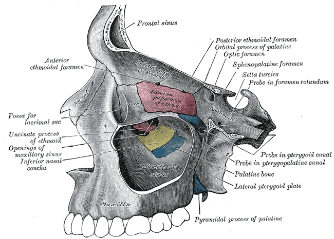
Gray's Anatomy (1918) plate, public domain
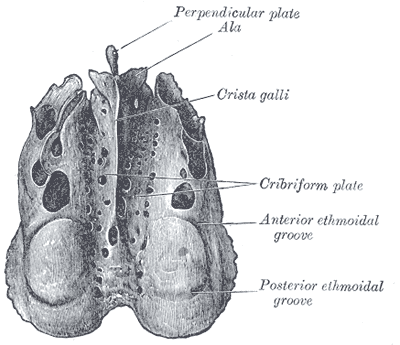
The bony orbit is the cone-shaped socket that holds and protects the eyeball. Seven bones contribute to each orbit: the frontal, sphenoid, zygomatic, maxilla, lacrimal, ethmoid and palatine. Openings at its back carry the optic nerve and the vessels and nerves of eye movement; its floor is thin and can give way in injuries.
Key facts
- Seven bones form each eye socket.
- The orbit protects the eye from all sides except the front.
- The optic canal at its tip carries the optic nerve to the brain.
- Its floor, formed largely by the maxilla, is thin and is the usual site of 'blowout' fractures.
- Fat cushions the eyeball inside the socket.
What can go wrong
A blow to the eye can fracture the thin orbital floor — a blowout fracture — sometimes trapping an eye muscle and causing double vision when looking up. The socket can also be affected by thyroid eye disease, in which the tissues inside it swell, and by infections spreading from the neighbouring sinuses.
Related procedures & tests
Related symptoms
Related muscles
Watch: related videos
Frequently asked questions
Why does the eye socket fracture instead of the eyeball bursting?
A blow compresses the soft contents of the socket and the pressure escapes through its weakest wall — the thin floor — which fractures. In effect the bone gives way first, which can protect the eyeball itself.
Which bones form the eye socket?
Seven: frontal above, zygomatic at the outer side, maxilla below, and the lacrimal, ethmoid, palatine and sphenoid bones making up the inner wall and the back of the socket.
Educational information only. This atlas explains normal anatomy and describes conditions in general terms; it is not medical advice and does not replace assessment by a qualified clinician. If you have symptoms that worry you, seek medical care.