- Physical Examination
- Surgical Examination
- Ophthalmology
- Clinical Skills
- Orthopedics
- Surgery Videos
- Laparoscopy
- Pediatrics
- Funny Videos
- Cardiothoracic Surgery
- Nursing Videos
- Plastic Surgery
- Otorhinolaryngology
- Histology and Histopathology
- Neurosurgery
- Dermatology
- Pediatric Surgery
- Urology
- Dentistry
- Oncology and Cancers
- Anatomy Videos
- Health and Fitness
- Radiology
- Anaesthesia
- Physical Therapy
- Pharmacology
- Interventional Radiology
- Cardiology
- Endocrinology
- Gynecology
- Emergency Medicine
- Psychiatry and Psychology
- Childbirth Videos
- General Medical Videos
- Nephrology
- Physiology
- Diet and Food Health
- Diabetes Mellitus
- Neurology
- Women Health
- Osteoporosis
- Gastroenterology
- Pulmonology
- Hematology
- Rheumatology
- Toxicology
- Nuclear Medicine
- Infectious Diseases
- Vascular Disease
- Reproductive Health
- Burns and Wound Healing
- Kidney Zone Podcast
- Other
Frontal Bone
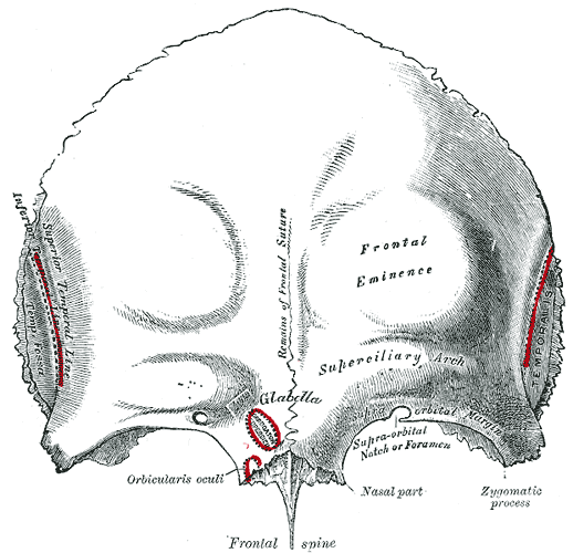
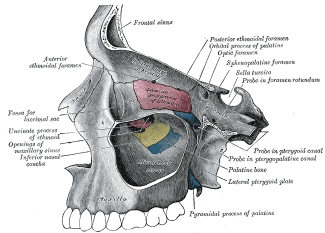
Gray's Anatomy (1918) plate, public domain
The frontal bone forms the forehead, the roof of the eye sockets and the front part of the skull vault. It is a single bone in adults, though babies are born with it in two halves that fuse during early childhood. Behind the brow ridges it contains the two frontal sinuses.
Key facts
- The frontal bone forms the forehead and the top of the eye sockets (orbits).
- It meets the parietal bones at the coronal suture across the top of the head.
- The raised brow ridges are called the superciliary arches; the smooth area between them is the glabella.
- It contains the frontal sinuses — air spaces that connect to the nose.
- At birth it is two bones joined by the metopic suture, which usually fuses by about age 8.
What can go wrong
The frontal bone can fracture in forehead injuries, sometimes denting inward. Its sinuses are a common site of sinusitis, felt as pressure or pain across the forehead. Very rarely, the childhood metopic suture fuses too early, changing the shape of the forehead — something checked at baby health visits.
Related procedures & tests
Related symptoms
Related muscles
Frequently asked questions
Is the frontal bone one bone or two?
Both, depending on age: babies have two frontal bones joined by the metopic suture, which normally fuses during childhood so that adults have a single frontal bone. In a small number of adults the suture persists harmlessly for life.
Why do forehead knocks raise a lump so quickly?
The skin of the forehead is tightly bound to the muscle and bone beneath, and the area has a rich blood supply, so even a minor bump can produce a noticeable swelling (a haematoma) within minutes.
Educational information only. This atlas explains normal anatomy and describes conditions in general terms; it is not medical advice and does not replace assessment by a qualified clinician. If you have symptoms that worry you, seek medical care.