- Physical Examination
- Surgical Examination
- Ophthalmology
- Clinical Skills
- Orthopedics
- Surgery Videos
- Laparoscopy
- Pediatrics
- Funny Videos
- Cardiothoracic Surgery
- Nursing Videos
- Plastic Surgery
- Otorhinolaryngology
- Histology and Histopathology
- Neurosurgery
- Dermatology
- Pediatric Surgery
- Urology
- Dentistry
- Oncology and Cancers
- Anatomy Videos
- Health and Fitness
- Radiology
- Anaesthesia
- Physical Therapy
- Pharmacology
- Interventional Radiology
- Cardiology
- Endocrinology
- Gynecology
- Emergency Medicine
- Psychiatry and Psychology
- Childbirth Videos
- General Medical Videos
- Nephrology
- Physiology
- Diet and Food Health
- Diabetes Mellitus
- Neurology
- Women Health
- Osteoporosis
- Gastroenterology
- Pulmonology
- Hematology
- Rheumatology
- Toxicology
- Nuclear Medicine
- Infectious Diseases
- Vascular Disease
- Reproductive Health
- Burns and Wound Healing
- Kidney Zone Podcast
- Other
First Metacarpal
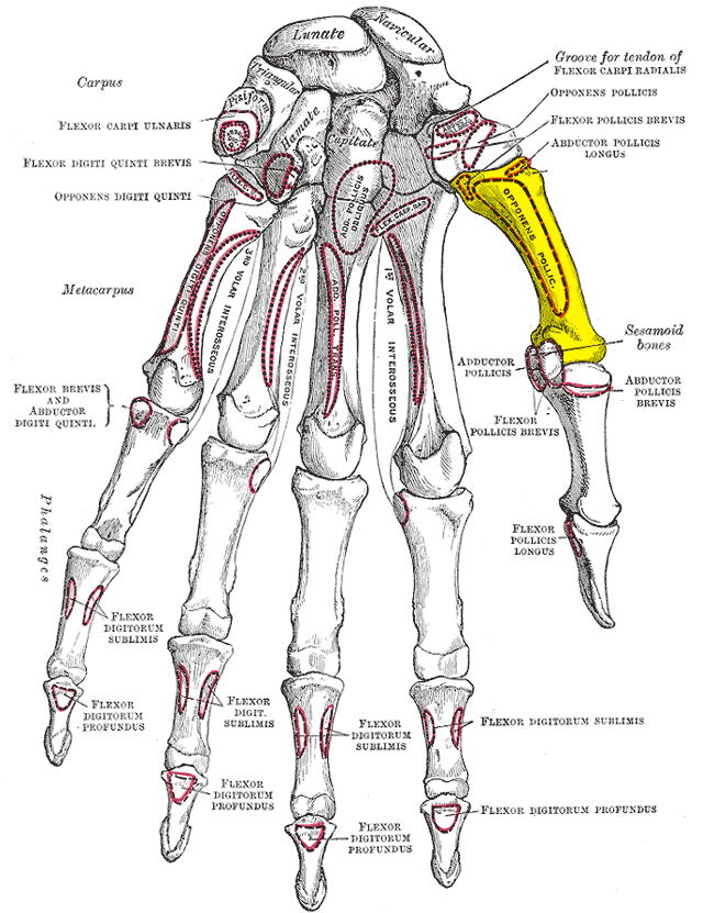
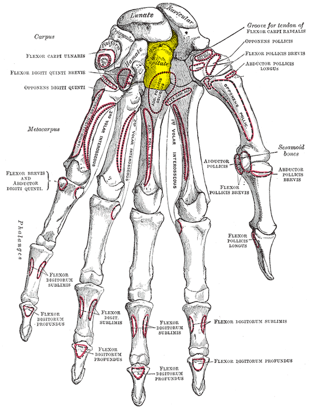
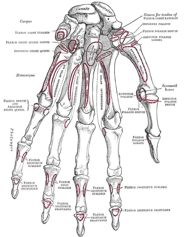
Gray's Anatomy (1918) plate, public domain
The first metacarpal is the thumb's palm bone — shorter and thicker than the others, and rotated about a quarter-turn so the thumb faces the fingers. Its saddle-shaped base joins the trapezium, and two small sesamoid bones sit under its head. This is the bone that makes the thumb a gripping tool rather than a fifth finger.
Key facts
- It is the shortest and most mobile metacarpal.
- Its rotation is what allows the thumb to oppose the fingers.
- Two sesamoid bones lie in the tendons beneath its head.
- The main thumb muscles (thenar muscles) act through it.
- Its base fracture (Bennett's fracture) is a classic hand injury.
What can go wrong
The first metacarpal's base can fracture in falls and fights; the Bennett fracture-dislocation, where a fragment stays in place while the rest of the bone dislocates, usually needs surgery to keep the thumb working. Arthritis at its saddle joint is the commonest arthritis in the hand.
Related procedures & tests
Related symptoms
Related muscles
Watch: related videos
Frequently asked questions
Why is the thumb's metacarpal set at an angle?
It is rotated roughly 90 degrees relative to the finger metacarpals, turning the thumb's plane of bending towards the palm. Without that rotation, the thumb could only wag side to side and pinch grip would be impossible.
What is a Bennett fracture?
A fracture-dislocation at the base of the thumb metacarpal: a small inner fragment stays with the trapezium while the rest of the bone is pulled out of place by the thumb's tendons. It is usually pinned or screwed in surgery.
Educational information only. This atlas explains normal anatomy and describes conditions in general terms; it is not medical advice and does not replace assessment by a qualified clinician. If you have symptoms that worry you, seek medical care.