- Physical Examination
- Surgical Examination
- Ophthalmology
- Clinical Skills
- Orthopedics
- Surgery Videos
- Laparoscopy
- Pediatrics
- Funny Videos
- Cardiothoracic Surgery
- Nursing Videos
- Plastic Surgery
- Otorhinolaryngology
- Histology and Histopathology
- Neurosurgery
- Dermatology
- Pediatric Surgery
- Urology
- Dentistry
- Oncology and Cancers
- Anatomy Videos
- Health and Fitness
- Radiology
- Anaesthesia
- Physical Therapy
- Pharmacology
- Interventional Radiology
- Cardiology
- Endocrinology
- Gynecology
- Emergency Medicine
- Psychiatry and Psychology
- Childbirth Videos
- General Medical Videos
- Nephrology
- Physiology
- Diet and Food Health
- Diabetes Mellitus
- Neurology
- Women Health
- Osteoporosis
- Gastroenterology
- Pulmonology
- Hematology
- Rheumatology
- Toxicology
- Nuclear Medicine
- Infectious Diseases
- Vascular Disease
- Reproductive Health
- Burns and Wound Healing
- Kidney Zone Podcast
- Other
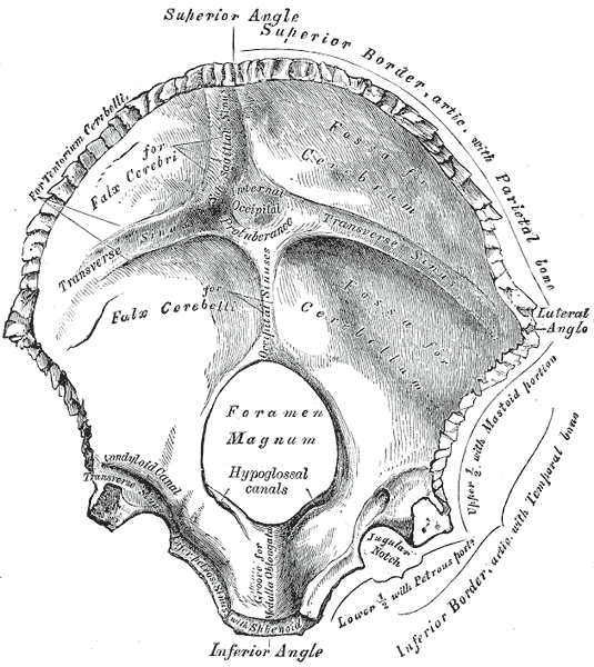
Occipital Bone
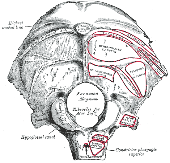
Gray's Anatomy (1918) plate, public domain
The occipital bone forms the back and base of the skull. It curves around the foramen magnum — the large opening where the spinal cord passes through to join the brain — and it has two smooth knobs (condyles) that rest on the first neck vertebra, letting you nod your head.
Key facts
- The occipital bone contains the foramen magnum, the largest opening in the skull.
- Its two condyles sit on the atlas (first neck vertebra) to form the nodding joint.
- Inside, it cradles the cerebellum, the part of the brain that coordinates movement.
- The bump at the back of the head (inion) is a landmark on this bone.
- Strong neck and back muscles attach along its curved nuchal lines.
What can go wrong
Injuries to the back of the head can fracture the occipital bone, and because the brainstem sits just inside it, such injuries are taken seriously. Some people are born with variations at the joint between this bone and the spine that narrow the space for the spinal cord and need specialist assessment.
Related procedures & tests
Related symptoms
Related muscles
Watch: related videos
Frequently asked questions
What passes through the hole in the occipital bone?
The foramen magnum transmits the spinal cord where it becomes the brainstem, the vertebral arteries that supply the brain, and parts of the accessory nerve. It is the main gateway between the skull and the spinal canal.
Which head movement does the occipital bone allow?
Nodding. The occipital condyles rock on the atlas below them, producing the yes movement of the head; turning the head side to side happens one joint lower.
Educational information only. This atlas explains normal anatomy and describes conditions in general terms; it is not medical advice and does not replace assessment by a qualified clinician. If you have symptoms that worry you, seek medical care.