- Physical Examination
- Surgical Examination
- Ophthalmology
- Clinical Skills
- Orthopedics
- Surgery Videos
- Laparoscopy
- Pediatrics
- Funny Videos
- Cardiothoracic Surgery
- Nursing Videos
- Plastic Surgery
- Otorhinolaryngology
- Histology and Histopathology
- Neurosurgery
- Dermatology
- Pediatric Surgery
- Urology
- Dentistry
- Oncology and Cancers
- Anatomy Videos
- Health and Fitness
- Radiology
- Anaesthesia
- Physical Therapy
- Pharmacology
- Interventional Radiology
- Cardiology
- Endocrinology
- Gynecology
- Emergency Medicine
- Psychiatry and Psychology
- Childbirth Videos
- General Medical Videos
- Nephrology
- Physiology
- Diet and Food Health
- Diabetes Mellitus
- Neurology
- Women Health
- Osteoporosis
- Gastroenterology
- Pulmonology
- Hematology
- Rheumatology
- Toxicology
- Nuclear Medicine
- Infectious Diseases
- Vascular Disease
- Reproductive Health
- Burns and Wound Healing
- Kidney Zone Podcast
- Other
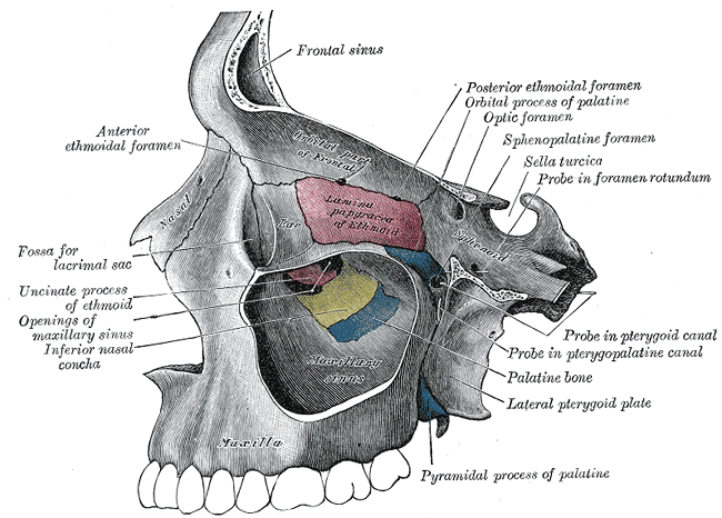
Palatine Bone
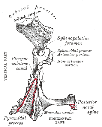
Gray's Anatomy (1918) plate, public domain
The palatine bones are two L-shaped bones at the back of the mouth. They form the back third of the hard palate (the roof of the mouth), part of the floor and side wall of the nose, and a small part of the floor of the eye socket. They are hidden deep inside the skull and rarely seen on the surface.
Key facts
- The palatine bones form the posterior third of the hard palate.
- They help separate the mouth from the nasal cavity.
- Each also contributes to the floor of the eye socket and the wall of the nose.
- Canals through them carry nerves and vessels to the palate.
- They are among the least visible bones of the skull in everyday examination.
What can go wrong
The palate matters most at birth: if the palatine bones and the tissues over them fail to fuse fully during development, a baby is born with a cleft palate — an opening between mouth and nose that affects feeding and speech and is repaired surgically in infancy. Later in life the palatine bones are seldom a problem on their own.
Frequently asked questions
What does the palatine bone separate?
Together with the maxilla, it forms the hard palate — the shelf of bone that separates the mouth below from the nasal cavity above, letting us breathe while we chew.
What is a cleft palate?
A gap in the roof of the mouth present from birth, where the palatal shelves did not fuse completely. It is usually repaired with surgery in the first year or so of life.
Educational information only. This atlas explains normal anatomy and describes conditions in general terms; it is not medical advice and does not replace assessment by a qualified clinician. If you have symptoms that worry you, seek medical care.