- Physical Examination
- Surgical Examination
- Ophthalmology
- Clinical Skills
- Orthopedics
- Surgery Videos
- Laparoscopy
- Pediatrics
- Funny Videos
- Cardiothoracic Surgery
- Nursing Videos
- Plastic Surgery
- Otorhinolaryngology
- Histology and Histopathology
- Neurosurgery
- Dermatology
- Pediatric Surgery
- Urology
- Dentistry
- Oncology and Cancers
- Anatomy Videos
- Health and Fitness
- Radiology
- Anaesthesia
- Physical Therapy
- Pharmacology
- Interventional Radiology
- Cardiology
- Endocrinology
- Gynecology
- Emergency Medicine
- Psychiatry and Psychology
- Childbirth Videos
- General Medical Videos
- Nephrology
- Physiology
- Diet and Food Health
- Diabetes Mellitus
- Neurology
- Women Health
- Osteoporosis
- Gastroenterology
- Pulmonology
- Hematology
- Rheumatology
- Toxicology
- Nuclear Medicine
- Infectious Diseases
- Vascular Disease
- Reproductive Health
- Burns and Wound Healing
- Kidney Zone Podcast
- Other
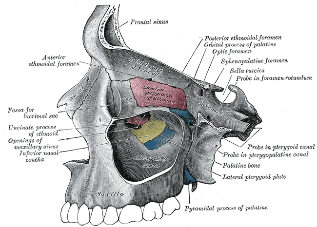
Paranasal Sinuses
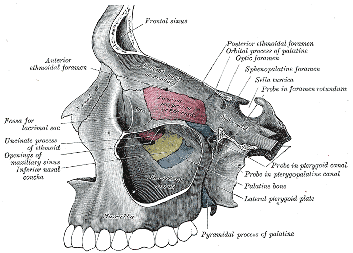
Gray's Anatomy (1918) plate, public domain
The paranasal sinuses are air-filled cavities inside the bones around the nose: the frontal sinuses in the forehead, the maxillary sinuses in the cheeks, the ethmoid cells between the eyes and the sphenoid sinuses deep behind the nose. They lighten the skull, add resonance to the voice and produce mucus that drains into the nose.
Key facts
- There are four pairs of paranasal sinuses, named after the bones that contain them.
- All of them open into the nasal cavity through small drainage channels.
- They are lined with the same mucus membrane as the nose.
- The maxillary sinuses are the largest; the sphenoid the deepest.
- They develop through childhood — the frontal sinuses appear last.
What can go wrong
Sinusitis — inflammation of the sinus lining, usually after a cold — is the commonest sinus problem, causing facial pressure, blockage and discharge. If drainage channels stay blocked, infections can recur. Persistent one-sided blockage or bleeding is checked promptly to exclude other causes.
Related symptoms
Watch: related videos
Frequently asked questions
What do the sinuses actually do?
They make the skull lighter than solid bone would be, give the voice some of its resonance, and their mucus lining humidifies inhaled air and traps particles, draining the mucus into the nose.
Why does sinusitis cause facial pain?
When the lining swells, the narrow drainage openings block and pressure builds inside the closed cavity; the inflamed lining itself is also pain-sensitive, producing the familiar ache over the cheeks or forehead.
Educational information only. This atlas explains normal anatomy and describes conditions in general terms; it is not medical advice and does not replace assessment by a qualified clinician. If you have symptoms that worry you, seek medical care.