- Physical Examination
- Surgical Examination
- Ophthalmology
- Clinical Skills
- Orthopedics
- Surgery Videos
- Laparoscopy
- Pediatrics
- Funny Videos
- Cardiothoracic Surgery
- Nursing Videos
- Plastic Surgery
- Otorhinolaryngology
- Histology and Histopathology
- Neurosurgery
- Dermatology
- Pediatric Surgery
- Urology
- Dentistry
- Oncology and Cancers
- Anatomy Videos
- Health and Fitness
- Radiology
- Anaesthesia
- Physical Therapy
- Pharmacology
- Interventional Radiology
- Cardiology
- Endocrinology
- Gynecology
- Emergency Medicine
- Psychiatry and Psychology
- Childbirth Videos
- General Medical Videos
- Nephrology
- Physiology
- Diet and Food Health
- Diabetes Mellitus
- Neurology
- Women Health
- Osteoporosis
- Gastroenterology
- Pulmonology
- Hematology
- Rheumatology
- Toxicology
- Nuclear Medicine
- Infectious Diseases
- Vascular Disease
- Reproductive Health
- Burns and Wound Healing
- Kidney Zone Podcast
- Other
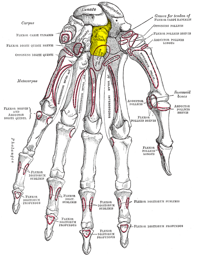
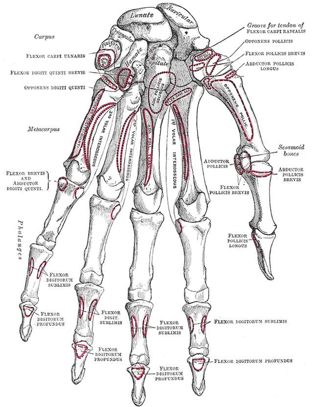
Second Metacarpal
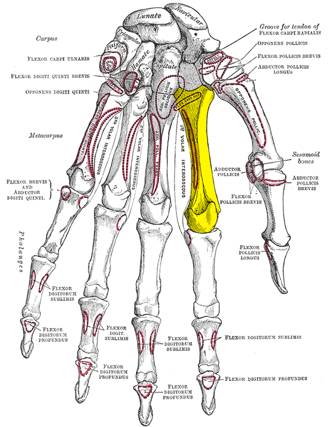
Gray's Anatomy (1918) plate, public domain
The second metacarpal is the index finger's palm bone, and the longest of the metacarpals. Its base is deeply wedged between the trapezium, trapezoid and capitate, making the index ray the least mobile of the hand — deliberately: it forms, with the middle ray, the rigid post against which the thumb pinches.
Key facts
- It has the longest metacarpal shaft.
- Its wedged base makes it the least mobile metacarpal.
- It forms the rigid post for pinch grip with the thumb.
- The first dorsal interosseous muscle — the bulk between thumb and index — originates from it.
- Its head is the index knuckle.
What can go wrong
Like other metacarpals it can fracture in crush injuries and falls; because the index ray is so stable, even modest displacement is noticed in pinch. The deep motor branch of the ulnar nerve and hardware placed for fractures are considerations in its surgery.
Related procedures & tests
Related symptoms
Related muscles
Frequently asked questions
Why does the index finger have the least mobile base?
Precision pinch needs a fixed target: the thumb moves, the index post stays. The wedge-shaped, tightly bound base of the second metacarpal provides that fixed post.
What is the muscle that bulges between thumb and index finger?
The first dorsal interosseous. It spreads the index finger away from the middle finger, and its bulk in the web space is supplied by a branch of the ulnar nerve — wasting there can be a sign of nerve problems.
Educational information only. This atlas explains normal anatomy and describes conditions in general terms; it is not medical advice and does not replace assessment by a qualified clinician. If you have symptoms that worry you, seek medical care.