- Physical Examination
- Surgical Examination
- Ophthalmology
- Clinical Skills
- Orthopedics
- Surgery Videos
- Laparoscopy
- Pediatrics
- Funny Videos
- Cardiothoracic Surgery
- Nursing Videos
- Plastic Surgery
- Otorhinolaryngology
- Histology and Histopathology
- Neurosurgery
- Dermatology
- Pediatric Surgery
- Urology
- Dentistry
- Oncology and Cancers
- Anatomy Videos
- Health and Fitness
- Radiology
- Anaesthesia
- Physical Therapy
- Pharmacology
- Interventional Radiology
- Cardiology
- Endocrinology
- Gynecology
- Emergency Medicine
- Psychiatry and Psychology
- Childbirth Videos
- General Medical Videos
- Nephrology
- Physiology
- Diet and Food Health
- Diabetes Mellitus
- Neurology
- Women Health
- Osteoporosis
- Gastroenterology
- Pulmonology
- Hematology
- Rheumatology
- Toxicology
- Nuclear Medicine
- Infectious Diseases
- Vascular Disease
- Reproductive Health
- Burns and Wound Healing
- Kidney Zone Podcast
- Other
Triquetrum
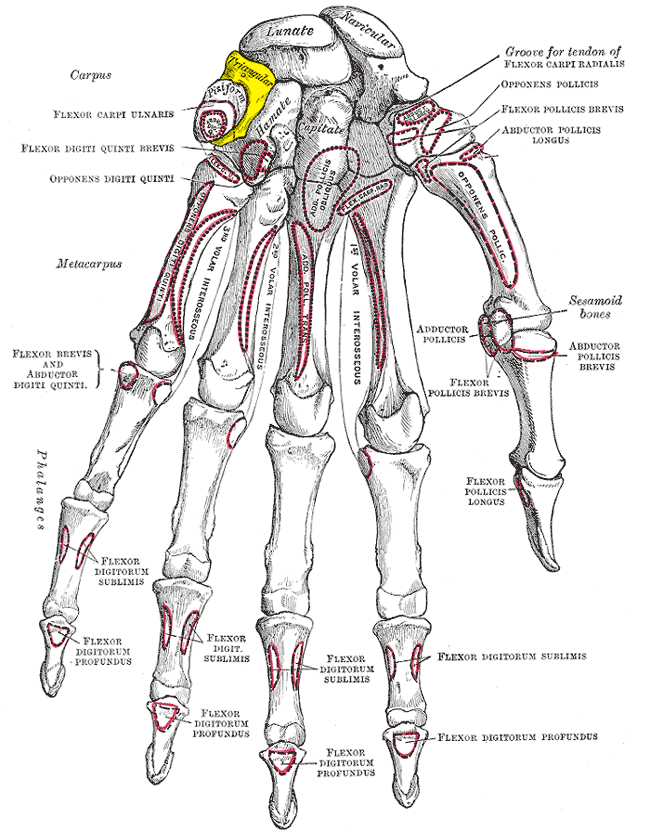
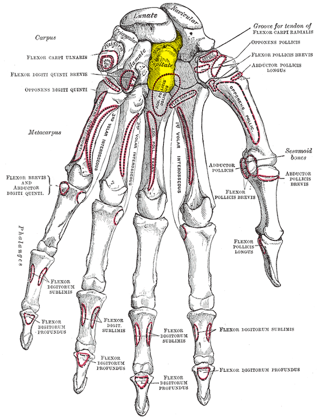
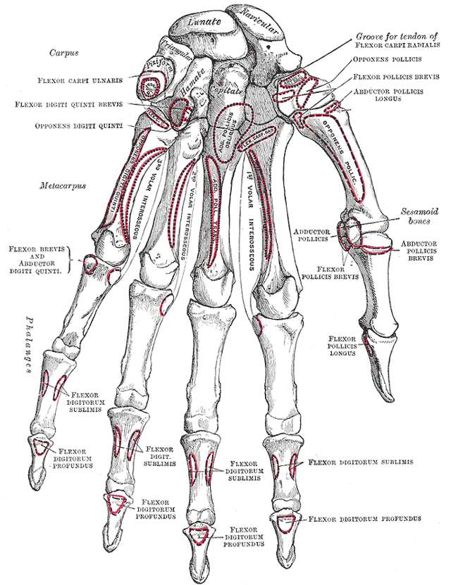
Gray's Anatomy (1918) plate, public domain
The triquetrum is the three-sided (pyramidal) carpal bone on the little-finger side of the wrist's near row. It meets the hamate and lunate, and carries the pea-shaped pisiform on its front surface. A pad of cartilage separates it from the ulna, so it does not meet the forearm bones directly.
Key facts
- The triquetrum is named for its three-cornered shape.
- It sits on the ulnar (little-finger) side of the wrist.
- The pisiform bone sits in a facet on its front surface.
- It does not touch the ulna — a cartilage disc fills the gap.
- Small chip fractures of its back surface are common in wrist sprains.
What can go wrong
The triquetrum is the second most often fractured carpal, usually a small chip flaked off its back surface when the wrist bends sharply backwards — often seen alongside sprains and usually treated with splinting. Pain on the ulnar side of the wrist has several causes, so persistent symptoms are examined carefully.
Related procedures & tests
Related symptoms
Related muscles
Frequently asked questions
What is the cartilage disc near the triquetrum?
Part of the triangular fibrocartilage complex (TFCC), a cushion and stabiliser between the ulna and the carpus. TFCC tears are a recognised cause of pain on the little-finger side of the wrist.
Are triquetrum chip fractures serious?
Usually not: most are small, stable flakes that heal with a few weeks of splinting. They matter mainly as a clue that the wrist ligaments were stressed.
Educational information only. This atlas explains normal anatomy and describes conditions in general terms; it is not medical advice and does not replace assessment by a qualified clinician. If you have symptoms that worry you, seek medical care.