- Physical Examination
- Surgical Examination
- Ophthalmology
- Clinical Skills
- Orthopedics
- Surgery Videos
- Laparoscopy
- Pediatrics
- Funny Videos
- Cardiothoracic Surgery
- Nursing Videos
- Plastic Surgery
- Otorhinolaryngology
- Histology and Histopathology
- Neurosurgery
- Dermatology
- Pediatric Surgery
- Urology
- Dentistry
- Oncology and Cancers
- Anatomy Videos
- Health and Fitness
- Radiology
- Anaesthesia
- Physical Therapy
- Pharmacology
- Interventional Radiology
- Cardiology
- Endocrinology
- Gynecology
- Emergency Medicine
- Psychiatry and Psychology
- Childbirth Videos
- General Medical Videos
- Nephrology
- Physiology
- Diet and Food Health
- Diabetes Mellitus
- Neurology
- Women Health
- Osteoporosis
- Gastroenterology
- Pulmonology
- Hematology
- Rheumatology
- Toxicology
- Nuclear Medicine
- Infectious Diseases
- Vascular Disease
- Reproductive Health
- Burns and Wound Healing
- Kidney Zone Podcast
- Other
Zygomatic Bone
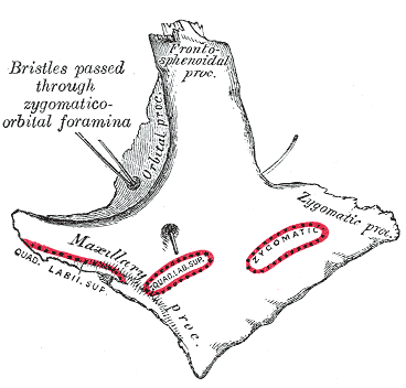
Gray's Anatomy (1918) plate, public domain
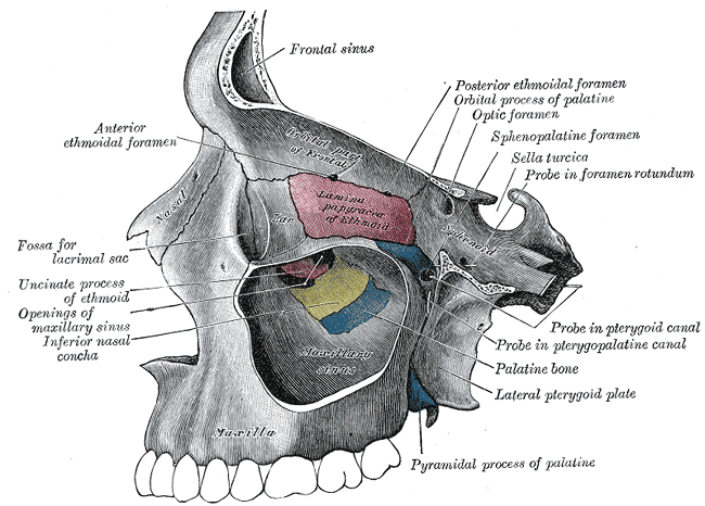
The zygomatic bones are the cheekbones. Each forms the prominence of the cheek, part of the lower and outer wall of the eye socket, and — reaching backwards — the zygomatic arch, the bony bridge you can feel under the skin in front of the ear. The masseter chewing muscle passes beneath this arch.
Key facts
- The zygomatic bone forms the point of the cheek.
- It helps form the floor and outer wall of the eye socket.
- Its backward process joins the temporal bone to form the zygomatic arch.
- The masseter muscle attaches to its lower edge and arch for chewing.
- It shapes the width of the face — high, prominent cheekbones are largely zygomatic bone.
What can go wrong
Cheekbone fractures are common in facial injuries from sport, falls and assaults; a depressed fracture can trap the masseter muscle or limit mouth opening, and fractures involving the socket can affect the eye. Many heal without surgery, but displaced ones may need an operation to restore the face's contour.
Related procedures & tests
Related muscles
Frequently asked questions
Can you feel the zygomatic arch?
Yes. Run a finger along the side of your face from the cheekbone towards the ear: the firm bony ridge under the skin is the zygomatic arch, formed by the zygomatic and temporal bones.
Why do cheekbone fractures matter for the eye?
The zygomatic bone forms part of the eye socket's floor and outer wall, so a fracture can change the socket's shape, and swelling or displacement can affect the position of the eyeball.
Educational information only. This atlas explains normal anatomy and describes conditions in general terms; it is not medical advice and does not replace assessment by a qualified clinician. If you have symptoms that worry you, seek medical care.