
- Physical Examination
- Surgical Examination
- Ophthalmology
- Clinical Skills
- Orthopedics
- Surgery Videos
- Laparoscopy
- Pediatrics
- Funny Videos
- Cardiothoracic Surgery
- Nursing Videos
- Plastic Surgery
- Otorhinolaryngology
- Histology and Histopathology
- Neurosurgery
- Dermatology
- Pediatric Surgery
- Urology
- Dentistry
- Oncology and Cancers
- Anatomy Videos
- Health and Fitness
- Radiology
- Anaesthesia
- Physical Therapy
- Pharmacology
- Interventional Radiology
- Cardiology
- Endocrinology
- Gynecology
- Emergency Medicine
- Psychiatry and Psychology
- Childbirth Videos
- General Medical Videos
- Nephrology
- Physiology
- Diet and Food Health
- Diabetes Mellitus
- Neurology
- Women Health
- Osteoporosis
- Gastroenterology
- Pulmonology
- Hematology
- Rheumatology
- Toxicology
- Nuclear Medicine
- Infectious Diseases
- Vascular Disease
- Reproductive Health
- Burns and Wound Healing
- Kidney Zone Podcast
- Other
Ischemia Patterns on an ECG
Ischemia patterns on an ECG — ST depression and T-wave changes — show heart muscle struggling for blood without the complete blockage of a STEMI. This page explains what these patterns look like, what they can mean, and when they are urgent, in plain language with videos.
✓ Medically reviewed by Dr. Mohamed Ibrahim, MD, PhD — physician-scientist · Last reviewed: November 2026
Watch: Ischemia Patterns on an ECG videos
 Heart Attack 3D Animation48,601 views
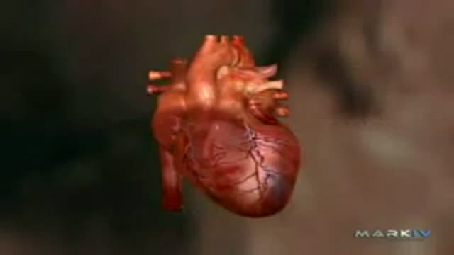 Acute Myocardial Infarction - Heart Attack30,283 views
 What happens during a Heart Attack?17,434 views
 Chronic Angina7,773 views
 Prinzmetal's Variant Angina4,635 views
 Microvascular Angina3,776 views
Guide
What it is
Ischemia means heart muscle is not getting enough oxygen-rich blood, usually because a coronary artery is narrowed by fatty deposits. Unlike a STEMI, the artery is not suddenly and completely blocked, so the ECG changes are subtler and reflect the inner layer of the heart wall struggling first. Ischemia may occur only during exertion or stress, when demand rises, or during unstable episodes at rest. The ECG is one of the fastest ways to detect it, especially when a tracing is captured while the person has symptoms.
What the tracing shows
The most common ischemia pattern is ST depression: the ST segment sits below the baseline in the leads facing the affected area. T waves may flatten or turn upside down (T-wave inversion). These changes often appear in neighbouring leads that view the same region, which helps locate the problem. Unlike STEMI, the changes can come and go with pain and may be absent at rest. Clinicians compare with older tracings, because long-standing identical changes are less worrying than new ones, and check blood tests for heart-muscle injury alongside.
What it can mean
Context decides how these patterns are handled. ST depression or T-wave inversion appearing with chest pain is treated as a likely acute coronary syndrome — the umbrella term for heart attack and unstable angina — until proven otherwise, with blood tests for heart-muscle injury and hospital assessment. The same changes on a routine tracing in someone who feels well prompt a planned work-up instead. Wellens syndrome deserves special mention: deeply inverted or two-peaked T waves in the front chest leads, in someone whose pain has settled, warn of a critical narrowing and need urgent specialist care, not a stress test.
When it is urgent
New ST depression or T-wave inversion with chest pain, breathlessness, sweating, or pain spreading to the arm or jaw is an emergency — call emergency services rather than driving yourself. Do not be reassured by pain easing: patterns like Wellens appear exactly when pain has settled, yet signal a dangerous narrowing. If ischemia changes are found on a routine ECG and you feel well, expect prompt follow-up — often same-week assessment, medicines to protect the heart, and further tests such as a stress test or angiogram.
Common questions
What is ischemia on an ECG?
Ischemia is heart muscle receiving too little blood and oxygen, usually because a coronary artery is narrowed. On an ECG it most often shows as ST-segment depression or T-wave inversion in the leads viewing the affected area.
What does ST depression mean?
ST depression means the ST segment sits below the baseline. It can reflect ischemia, but it also occurs with heart strain, some medicines such as digoxin, and low potassium, so it is read in context.
What is Wellens syndrome?
Wellens syndrome is a pattern of deeply inverted or two-peaked T waves in the front chest leads, seen after chest pain has settled. It warns of a critical narrowing of a major artery and needs urgent specialist care.
Can an ECG be normal during ischemia?
Yes. Ischemia can be present with a normal resting ECG, especially when blood flow is adequate at rest. That is why stress tests and symptom assessment exist, and why chest pain is never dismissed on one normal tracing.
More in Heart Attack & Ischemia Patterns
Related
Sources
- Heart Attack (MedlinePlus)
- Heart attack (NHS)
- About Heart Disease (CDC)
- Electrocardiogram (ECG) test (MedlinePlus)
This guide is general information, not medical advice, and does not replace advice from your own clinician. ECG patterns should always be interpreted in context by a trained professional. If you have chest pain, severe breathlessness, fainting or a racing heartbeat that will not settle, seek urgent medical care. Ischemia patterns on an ECG need urgent clinical assessment when new; this page explains patterns and is not a self-diagnosis tool.