
- Physical Examination
- Surgical Examination
- Ophthalmology
- Clinical Skills
- Orthopedics
- Surgery Videos
- Laparoscopy
- Pediatrics
- Funny Videos
- Cardiothoracic Surgery
- Nursing Videos
- Plastic Surgery
- Otorhinolaryngology
- Histology and Histopathology
- Neurosurgery
- Dermatology
- Pediatric Surgery
- Urology
- Dentistry
- Oncology and Cancers
- Anatomy Videos
- Health and Fitness
- Radiology
- Anaesthesia
- Physical Therapy
- Pharmacology
- Interventional Radiology
- Cardiology
- Endocrinology
- Gynecology
- Emergency Medicine
- Psychiatry and Psychology
- Childbirth Videos
- General Medical Videos
- Nephrology
- Physiology
- Diet and Food Health
- Diabetes Mellitus
- Neurology
- Women Health
- Osteoporosis
- Gastroenterology
- Pulmonology
- Hematology
- Rheumatology
- Toxicology
- Nuclear Medicine
- Infectious Diseases
- Vascular Disease
- Reproductive Health
- Burns and Wound Healing
- Kidney Zone Podcast
- Other
The 12 Leads
The 12 leads of an ECG are 12 different electrical viewpoints of the same heartbeat, recorded from 10 electrodes on the chest, arms, and legs. This page explains what each lead looks at and why having several views matters, in plain language with videos.
✓ Medically reviewed by Dr. Mohamed Ibrahim, MD, PhD — physician-scientist · Last reviewed: November 2026
Watch: The 12 Leads videos
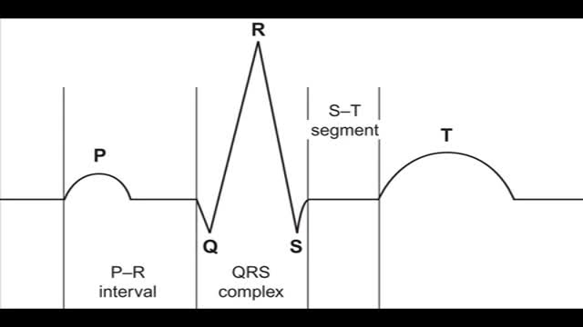 Reading the 12-lead ECG19,707 views
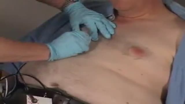 ECG Leads Placement12,140 views
 ECG Electrodes placement11,406 views
Guide
What it is
Each lead compares the electrical signal between two points, or between one point and an average of others, and draws the heartbeat from that angle. The six limb leads — I, II, III, aVR, aVL, and aVF — are calculated from electrodes on the arms and legs. The six chest leads — V1 to V6 — come from six electrodes placed in a line across the chest. Because the heart sits at an angle in the chest, no single lead sees the whole organ, so the 12 views are read together as one picture.
What the tracing shows
On the printout, each lead shows the same few beats drawn from its own angle, so a wave that points upward in one lead may point downward in another — that is normal and expected. Lead II usually gives the clearest view of the rhythm and is often used for continuous monitoring. The chest leads change gradually from V1 to V6 as their viewpoint moves around the heart. Clinicians check that the pattern of change is smooth, because a sudden change in one area can point to where heart muscle is injured or strained.
What it can mean
Grouped leads view the same region of heart muscle, so a change that appears in one group but not others helps locate a problem. The lower wall of the heart is viewed mainly by leads II, III, and aVF; the front wall by the middle chest leads; and the side wall by I, aVL, and the later chest leads. This is why a heart attack pattern is usually required in at least two neighbouring leads before it is trusted: a real regional problem should be visible from more than one nearby viewpoint.
When it is urgent
Lead placement matters: electrodes put in the wrong position can create patterns that look like disease on a healthy heart, or hide real changes. If a tracing looks unexpectedly abnormal, the team may check the electrode positions and repeat the recording before acting on it. Chest pain with a worrying pattern in neighbouring leads is treated as an emergency. Extra leads are sometimes added — for example on the back or the right side of the chest — when doctors need a view the standard 12 do not give.
Common questions
Why are there 12 leads but only 10 electrodes?
The 12 leads are calculated views, not wires. Four limb electrodes produce six limb-lead views by comparing signals in different combinations, and six chest electrodes each give one chest-lead view.
What do the chest leads V1 to V6 show?
The chest leads view the front of the heart as their positions move from the right side of the breastbone around to the left side of the chest. They are especially useful for seeing the front wall of the main pumping chamber.
Can leads be placed wrongly?
Yes. Swapped arm electrodes or chest electrodes placed too high or too low can change the tracing and imitate disease. This is a common reason for repeating an ECG before making decisions.
What does it mean when a change is in neighbouring leads?
Neighbouring leads view the same region of the heart, so a change appearing in two or more of them is more likely to be real and helps pinpoint which part of the heart muscle is affected.
More in Basics
Related
Sources
- Electrocardiogram (ECG) test (MedlinePlus)
- Electrocardiogram (MedlinePlus Medical Encyclopedia)
- ECG (electrocardiogram) (British Heart Foundation)
- Electrocardiogram (ECG) (NHS)
This guide is general information, not medical advice, and does not replace advice from your own clinician. ECG patterns should always be interpreted in context by a trained professional. If you have chest pain, severe breathlessness, fainting or a racing heartbeat that will not settle, seek urgent medical care. Lead positions and groupings are described in general terms; formal ECG interpretation is done by trained clinicians using the full tracing.