
- Physical Examination
- Surgical Examination
- Ophthalmology
- Clinical Skills
- Orthopedics
- Surgery Videos
- Laparoscopy
- Pediatrics
- Funny Videos
- Cardiothoracic Surgery
- Nursing Videos
- Plastic Surgery
- Otorhinolaryngology
- Histology and Histopathology
- Neurosurgery
- Dermatology
- Pediatric Surgery
- Urology
- Dentistry
- Oncology and Cancers
- Anatomy Videos
- Health and Fitness
- Radiology
- Anaesthesia
- Physical Therapy
- Pharmacology
- Interventional Radiology
- Cardiology
- Endocrinology
- Gynecology
- Emergency Medicine
- Psychiatry and Psychology
- Childbirth Videos
- General Medical Videos
- Nephrology
- Physiology
- Diet and Food Health
- Diabetes Mellitus
- Neurology
- Women Health
- Osteoporosis
- Gastroenterology
- Pulmonology
- Hematology
- Rheumatology
- Toxicology
- Nuclear Medicine
- Infectious Diseases
- Vascular Disease
- Reproductive Health
- Burns and Wound Healing
- Kidney Zone Podcast
- Other
Reading an ECG Step by Step
Reading an ECG step by step means checking the same features in the same order every time: rate, rhythm, intervals, and the shape of the waves. This page walks through that routine in plain language, with videos, so a tracing feels less like a mystery and more like a checklist.
✓ Medically reviewed by Dr. Mohamed Ibrahim, MD, PhD — physician-scientist · Last reviewed: November 2026
Watch: Reading an ECG Step by Step videos
 How to read ECG Part 176,124 views
 How to read ECG Part 229,723 views
 How to Read ECG Part 321,838 views
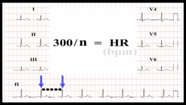 ECG Interpretation20,810 views
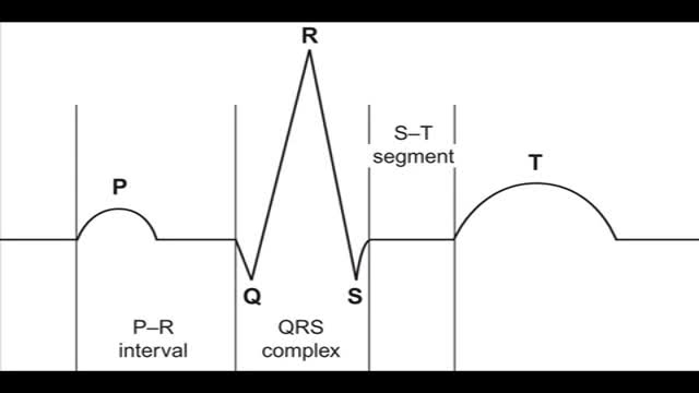 Reading the 12-lead ECG19,707 views
 EKG/ECG Interpretation Explained Clearly1,667 views
Guide
What it is
Clinicians read ECGs systematically rather than glancing at the overall shape. The routine on this page is the plain-language version of that approach. It works because each step answers a separate question: how fast, how regular, how the signal travels, and how the muscle responds. You will not become an expert from one page — formal interpretation takes training and the patient's full story — but you will understand what each line of an ECG report means and why it is there.
What the tracing shows
Step one is rate: at the standard paper speed, count the large squares between two neighbouring QRS spikes and divide 300 by that number to estimate beats per minute; a normal resting rate is 60 to 100. Step two is rhythm: check the spacing between beats is even and that a P wave comes before every QRS. Step three is the intervals: PR, QRS width, and QTc against their usual ranges. Step four is the axis and shapes: the general direction of the electrical signal — normally pointing down and to the left — and whether the ST segments sit level with the baseline.
What it can mean
Each step rules problems in or out. An uneven rhythm with no clear P waves suggests atrial fibrillation; a very wide QRS suggests a conduction block or a ventricular rhythm; a raised ST segment in neighbouring leads suggests a heart attack pattern. Reading in order also protects against false alarms: a single odd-looking beat or one noisy lead is common and usually harmless. The final step is always comparison — a pattern that is new for that person matters far more than one that has been present, unchanged, for years.
When it is urgent
Some findings should never wait for a routine report: ST elevation with chest pain, a very fast or very slow rate with fainting or breathlessness, or a completely irregular rhythm in someone who feels unwell. In hospital, ECGs with these patterns are escalated immediately. If you receive a result mentioning a long QT, heart block, or ST changes, expect follow-up such as a repeat tracing, blood tests, a medicine review, or referral to a heart specialist — and seek urgent help at once if you develop chest pain, severe palpitations, or fainting.
Common questions
How is heart rate calculated from an ECG?
A common method is to divide 300 by the number of large grid squares between two neighbouring QRS spikes. Five large squares, for example, gives about 60 beats per minute, and the method works best when the rhythm is regular.
What is the normal heart rate on an ECG?
A normal resting heart rate is 60 to 100 beats per minute. Rates below 60 are called bradycardia and above 100 tachycardia, though fit people often rest below 60 and rates rise normally with exercise, pain, or fever.
What is the heart's axis, in simple terms?
The axis is the overall direction the electrical signal travels through the heart, which normally points downwards and towards the left. A shifted axis can hint at chamber enlargement or a conduction problem, and is one clue among many.
Why is my ECG compared with an old one?
Comparison separates new problems from long-standing patterns. Many ECG variations are normal for the individual and unchanged for years, while a change from a previous tracing can be the first sign of new disease.
More in Basics
Related
Sources
- Electrocardiogram (MedlinePlus Medical Encyclopedia)
- Electrocardiogram (ECG) test (MedlinePlus)
- ECG (electrocardiogram) (British Heart Foundation)
- Electrocardiogram (ECG) (NHS)
This guide is general information, not medical advice, and does not replace advice from your own clinician. ECG patterns should always be interpreted in context by a trained professional. If you have chest pain, severe breathlessness, fainting or a racing heartbeat that will not settle, seek urgent medical care. This routine explains how ECG reports are structured; it is not a substitute for formal interpretation by a trained clinician.