
- Physical Examination
- Surgical Examination
- Ophthalmology
- Clinical Skills
- Orthopedics
- Surgery Videos
- Laparoscopy
- Pediatrics
- Funny Videos
- Cardiothoracic Surgery
- Nursing Videos
- Plastic Surgery
- Otorhinolaryngology
- Histology and Histopathology
- Neurosurgery
- Dermatology
- Pediatric Surgery
- Urology
- Dentistry
- Oncology and Cancers
- Anatomy Videos
- Health and Fitness
- Radiology
- Anaesthesia
- Physical Therapy
- Pharmacology
- Interventional Radiology
- Cardiology
- Endocrinology
- Gynecology
- Emergency Medicine
- Psychiatry and Psychology
- Childbirth Videos
- General Medical Videos
- Nephrology
- Physiology
- Diet and Food Health
- Diabetes Mellitus
- Neurology
- Women Health
- Osteoporosis
- Gastroenterology
- Pulmonology
- Hematology
- Rheumatology
- Toxicology
- Nuclear Medicine
- Infectious Diseases
- Vascular Disease
- Reproductive Health
- Burns and Wound Healing
- Kidney Zone Podcast
- Other
Waves and Intervals
The waves and intervals of an ECG are the named parts of each heartbeat's tracing: the P wave, the QRS complex, the T wave, and the timed gaps between them. This page explains what each part represents and the measurements usually considered normal, in plain language with videos.
✓ Medically reviewed by Dr. Mohamed Ibrahim, MD, PhD — physician-scientist · Last reviewed: November 2026
Watch: Waves and Intervals videos
 How to read ECG Part 176,124 views
 How to read ECG Part 229,723 views
 How to Read ECG Part 321,838 views
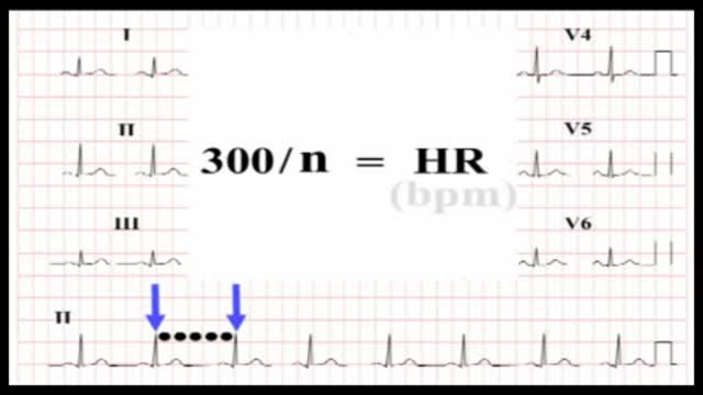 ECG Interpretation20,810 views
 The basics of the ECG17,257 views
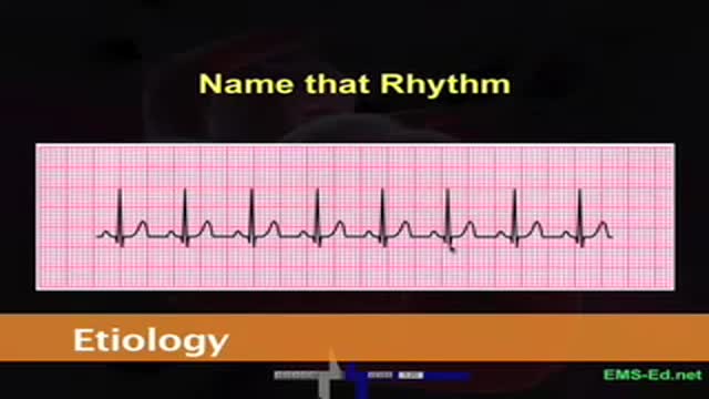 ECG part 25,062 views
Guide
What it is
Each heartbeat draws the same sequence. First comes the small, rounded P wave as the two upper chambers (atria) activate. After a short flat pause, the tall QRS complex appears as the two lower chambers (ventricles) activate — its sharp spike is the easiest part to spot. The tracing then returns to baseline before the rounded T wave, which shows the ventricles electrically resetting, ready for the next beat. Sometimes a small extra bump, a U wave, follows the T wave. Learning this sequence is the first step in reading any tracing.
What the tracing shows
ECGs are printed on a grid that measures time and size. At the standard speed, each small square is 1 millimetre and represents 0.04 seconds, and each large square of five small ones represents 0.20 seconds. The PR interval, from the start of the P wave to the start of the QRS, usually measures 0.12 to 0.20 seconds. The QRS itself is normally narrow — under 0.12 seconds. The QT interval runs from the start of the QRS to the end of the T wave and is corrected for heart rate (QTc), because it naturally shortens as the heart speeds up.
What it can mean
Each part points to a different part of the heart's electrical system. A long PR interval suggests delay at the junction between the chambers, the AV node. A wide QRS suggests the signal took a slower route through the ventricles, as in bundle branch block or a rhythm starting in the ventricles themselves. A long QTc points to slow electrical recovery, which can come from inherited conditions, low blood salts, or many medicines. Changes in the T wave and the ST segment — the short stretch between the QRS and the T wave — can reflect strain, low blood flow, or blood-salt problems.
When it is urgent
Some wave and interval changes need same-day or emergency assessment: a markedly long QT interval, a very wide QRS, or an ST segment that is newly raised or deeply pressed down, especially with chest pain, fainting, or severe palpitations. Because single measurements can mislead, clinicians measure intervals across several beats and leads, and compare with older tracings. If you are told an interval is abnormal, ask what is being compared and whether a repeat tracing, blood tests, or a medicine review is needed — those are the usual next steps.
Common questions
What is a normal PR interval?
A normal PR interval is usually 0.12 to 0.20 seconds — about three to five small squares on standard ECG paper. Longer than 0.20 seconds means the signal is delayed at the AV node, called first-degree heart block.
How wide should the QRS complex be?
The QRS complex is normally under 0.12 seconds wide. A QRS of 0.12 seconds or more is called wide and suggests the signal spread through the ventricles by a slower route, as in bundle branch block.
What is the QTc and why is it corrected?
The QT interval naturally gets shorter as the heart beats faster, so it is corrected for rate to give the QTc, which allows fair comparison. Cut-offs commonly used for a prolonged QTc are above 450 milliseconds in men and above 470 milliseconds in women.
What is the ST segment?
The ST segment is the short, usually flat stretch between the end of the QRS complex and the start of the T wave. It is watched closely because it rises during many heart attacks and dips when heart muscle is short of blood.
More in Basics
Related
Sources
- Electrocardiogram (MedlinePlus Medical Encyclopedia)
- Electrocardiogram (ECG) test (MedlinePlus)
- Long QT syndrome (NHS)
- ECG (electrocardiogram) (British Heart Foundation)
This guide is general information, not medical advice, and does not replace advice from your own clinician. ECG patterns should always be interpreted in context by a trained professional. If you have chest pain, severe breathlessness, fainting or a racing heartbeat that will not settle, seek urgent medical care. Interval ranges quoted here are the commonly used adult reference values; exact cut-offs vary slightly between guidelines and are applied by clinicians in context.