
- Physical Examination
- Surgical Examination
- Ophthalmology
- Clinical Skills
- Orthopedics
- Surgery Videos
- Laparoscopy
- Pediatrics
- Funny Videos
- Cardiothoracic Surgery
- Nursing Videos
- Plastic Surgery
- Otorhinolaryngology
- Histology and Histopathology
- Neurosurgery
- Dermatology
- Pediatric Surgery
- Urology
- Dentistry
- Oncology and Cancers
- Anatomy Videos
- Health and Fitness
- Radiology
- Anaesthesia
- Physical Therapy
- Pharmacology
- Interventional Radiology
- Cardiology
- Endocrinology
- Gynecology
- Emergency Medicine
- Psychiatry and Psychology
- Childbirth Videos
- General Medical Videos
- Nephrology
- Physiology
- Diet and Food Health
- Diabetes Mellitus
- Neurology
- Women Health
- Osteoporosis
- Gastroenterology
- Pulmonology
- Hematology
- Rheumatology
- Toxicology
- Nuclear Medicine
- Infectious Diseases
- Vascular Disease
- Reproductive Health
- Burns and Wound Healing
- Kidney Zone Podcast
- Other
Chest X-ray
A chest X-ray is the most common imaging test in medicine — a single quick picture of the lungs, heart, and chest bones. This page explains what it shows, why it is ordered, and how to make sense of the words in the report, in plain language with videos.
✓ Medically reviewed by Dr. Mohamed Ibrahim, MD, PhD — physician-scientist · Last reviewed: November 2026
Watch: Chest X-ray videos
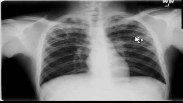 Chest x-ray --Tuberculosis30,298 views
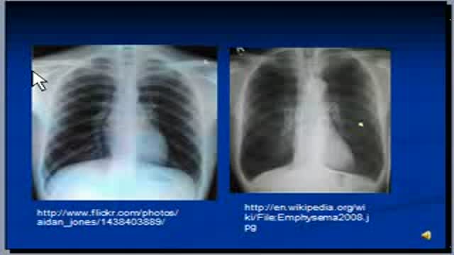 Chest x-ray interpretation -- COPD and Emphysema27,260 views
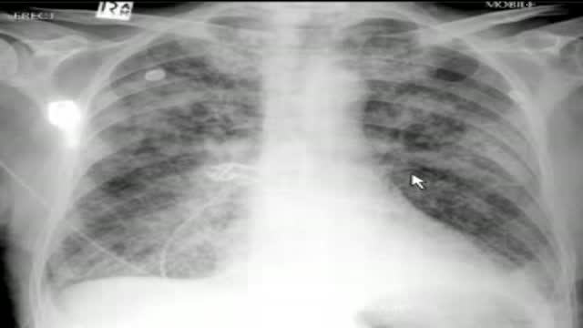 chest x-ray, pulmonary edema23,354 views
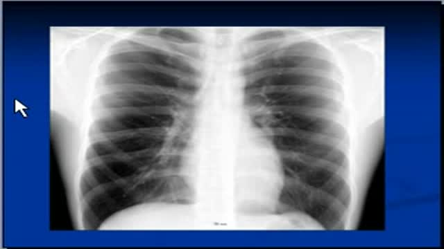 Chest x-ray --Silhouette sign22,186 views
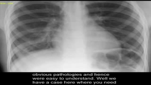 Pneumonia, on chest x-ray.21,247 views
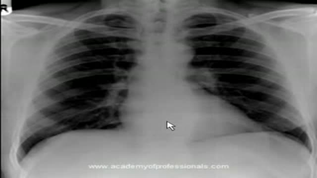 chest x-ray, artificial aortic and mitral valves19,960 views
Guide
What it is
For a standard chest X-ray you stand with your chest against a detector plate, take a deep breath in, and hold it for a few seconds while the picture is taken. A second picture from the side is usually added. If you are too unwell to stand, the picture can be taken sitting up or lying in bed, which gives a slightly less clear image. The whole appointment normally takes only a few minutes, and there is no recovery time — you can go straight back to your day or ward afterwards.
What it shows
Because air, soft tissue, and bone absorb X-rays differently, the chest is well suited to this test. Doctors look at whether the lungs are fully inflated and clear, whether fluid has collected around them (an effusion), whether air has leaked outside a lung (a collapsed lung or pneumothorax), and whether the heart outline is enlarged. The test also checks the position of tubes and lines in hospital patients, and the ribs and spine for obvious breaks. It cannot show fine detail inside the lung tissue, small early cancers reliably, or the heart's valves and muscle movement — those need CT or ultrasound of the heart.
How to prepare
There is nothing to eat, drink, or stop before a chest X-ray. You will undress from the waist up and wear a gown, and remove necklaces and any metal over the chest, because metal creates a bright white mark that can mimic or hide a problem. The radiographer will position you and ask you to breathe in deeply and hold still — a sharp, full breath spreads the lungs out and makes the picture much easier to read. Tell the team if you might be pregnant, or if you cannot stand, so they can adapt the examination.
What the report words mean
Chest X-ray reports use a shared vocabulary. 'Consolidation' means a patch of lung has filled with fluid or pus instead of air — most often pneumonia. An 'effusion' is fluid collected around the lung. 'Cardiomegaly' means the heart outline looks enlarged. 'Atelectasis' is a small collapsed, airless area, common after surgery. 'No focal consolidation' is a reassuring phrase meaning no pneumonia-like patch was seen. Words like 'opacity' simply describe a whiter patch and are not a diagnosis by themselves — the ordering clinician interprets them alongside your symptoms.
Common questions
Why do I have to hold my breath?
Holding a deep breath in keeps the lungs still and fully expanded for the few seconds of the exposure. A moving or half-empty lung looks blurred and can hide small problems or imitate them. The radiographer will tell you exactly when to breathe in, hold, and breathe normally again.
Can a chest X-ray find lung cancer?
A chest X-ray can show larger lung tumours and is often the first test that raises suspicion, but it can miss small or hidden cancers, especially behind the heart or ribs. If cancer is a concern, a CT scan is the more reliable test, and a normal chest X-ray does not completely exclude it.
How quickly will I get the result?
In an emergency department the ordering doctor usually reviews the image the same day, often within hours. For routine outpatient requests, a radiologist's written report is typically sent to your clinician within days. If something urgent is seen, the imaging team contacts the ordering clinician directly rather than waiting for routine reporting.
Is a chest X-ray safe in pregnancy?
The radiation dose from a chest X-ray is very small and the beam is aimed at the chest, away from the abdomen, which can also be shielded. It is still important to tell the team you are pregnant or might be, so they can confirm the test is needed and take any extra precautions.
More in X-ray & Fluoroscopy
Related
Sources
- Chest X-ray (MedlinePlus Medical Encyclopedia)
- Chest X-ray (Radiography — Chest) (RadiologyInfo.org (RSNA/ACR))
- X-ray (NHS)
This guide is general information, not medical advice, and does not replace advice from your own clinician. How a scan is done, and whether contrast is used, depends on your own medical history — always follow the instructions of the team performing your scan, and tell them about kidney problems, allergies, implants, and the medicines you take. A normal chest X-ray does not rule out every lung or heart problem; ongoing symptoms should always be reviewed by a clinician.