
- Physical Examination
- Surgical Examination
- Ophthalmology
- Clinical Skills
- Orthopedics
- Surgery Videos
- Laparoscopy
- Pediatrics
- Funny Videos
- Cardiothoracic Surgery
- Nursing Videos
- Plastic Surgery
- Otorhinolaryngology
- Histology and Histopathology
- Neurosurgery
- Dermatology
- Pediatric Surgery
- Urology
- Dentistry
- Oncology and Cancers
- Anatomy Videos
- Health and Fitness
- Radiology
- Anaesthesia
- Physical Therapy
- Pharmacology
- Interventional Radiology
- Cardiology
- Endocrinology
- Gynecology
- Emergency Medicine
- Psychiatry and Psychology
- Childbirth Videos
- General Medical Videos
- Nephrology
- Physiology
- Diet and Food Health
- Diabetes Mellitus
- Neurology
- Women Health
- Osteoporosis
- Gastroenterology
- Pulmonology
- Hematology
- Rheumatology
- Toxicology
- Nuclear Medicine
- Infectious Diseases
- Vascular Disease
- Reproductive Health
- Burns and Wound Healing
- Kidney Zone Podcast
- Other
MRI scan — knee
Category: Imaging & Scans · All imaging & scans
What it is
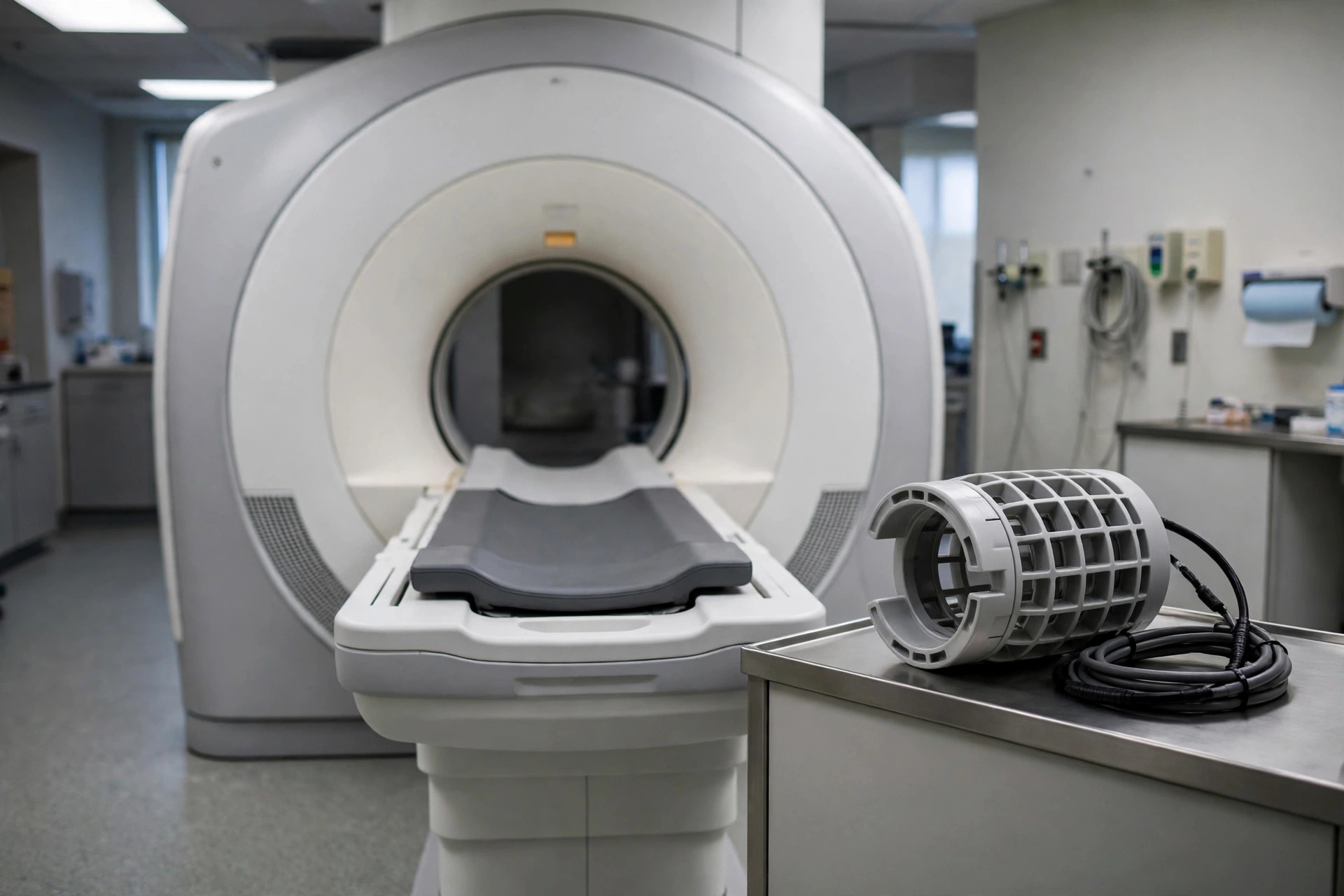
An MRI scan of the knee uses a magnet and radio waves to produce detailed pictures of the cartilage, ligaments, tendons and bones inside the knee joint.
Why it is done
It is often used after a knee injury, for ongoing knee pain, swelling or locking, or when a torn ligament or cartilage problem is suspected and the result would change treatment.
How to prepare
There is usually no special preparation beyond the standard MRI metal-safety checks. You can usually eat and drink normally.
What happens during the procedure
You lie on the table with your leg in a padded frame (coil) and only your leg goes into the scanner opening for most knee scans. It is noisy and takes about 20 to 30 minutes; keeping the leg still is important.
Afterwards — recovery & results
You can return to normal activities immediately. A radiologist reports the scan and your doctor will discuss the findings and next steps with you.
Risks & considerations
MRI has no ionising radiation. The magnet makes it unsuitable for some implants, which is why screening questions are asked. Lying still with an uncomfortable knee can be the hardest part for some people.
Related guides on MedicalVideos.com
Frequently asked questions
Will the scan decide if I need an operation?
It is one piece of the picture. Surgeons decide using your symptoms, the examination and the scan together — some tears found on MRI are managed without surgery.
Can I move during the scan?
Small movements blur the pictures, so the team will make you comfortable with cushions first. If you become uncomfortable, you can speak to them through the intercom.
Related procedures — Imaging & Scans
Browse the M section of the A–Z list or return to the Procedures & Tests hub.