
- Physical Examination
- Surgical Examination
- Ophthalmology
- Clinical Skills
- Orthopedics
- Surgery Videos
- Laparoscopy
- Pediatrics
- Funny Videos
- Cardiothoracic Surgery
- Nursing Videos
- Plastic Surgery
- Otorhinolaryngology
- Histology and Histopathology
- Neurosurgery
- Dermatology
- Pediatric Surgery
- Urology
- Dentistry
- Oncology and Cancers
- Anatomy Videos
- Health and Fitness
- Radiology
- Anaesthesia
- Physical Therapy
- Pharmacology
- Interventional Radiology
- Cardiology
- Endocrinology
- Gynecology
- Emergency Medicine
- Psychiatry and Psychology
- Childbirth Videos
- General Medical Videos
- Nephrology
- Physiology
- Diet and Food Health
- Diabetes Mellitus
- Neurology
- Women Health
- Osteoporosis
- Gastroenterology
- Pulmonology
- Hematology
- Rheumatology
- Toxicology
- Nuclear Medicine
- Infectious Diseases
- Vascular Disease
- Reproductive Health
- Burns and Wound Healing
- Kidney Zone Podcast
- Other
Fluoroscopy (real-time X-ray)
Category: Imaging & Scans · All imaging & scans
What it is
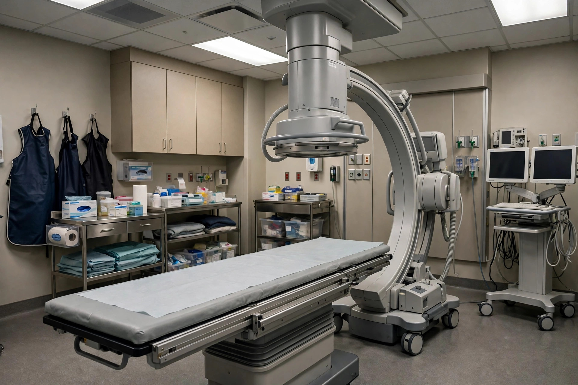
Fluoroscopy is a moving X-ray picture — like an X-ray video — that lets doctors watch structures and instruments in real time. It is used to guide many procedures and to watch swallowing, bowel movement and joint movement as they happen.
Why it is done
Common uses include barium studies of the digestive tract, guiding catheters and injections, checking the position of tubes and lines, and some bladder, womb and joint examinations.
How to prepare
Preparation depends entirely on the examination being done and your appointment letter will explain. Tell the team if you might be pregnant.
What happens during the procedure
You lie or stand by an X-ray table while the camera moves around you. The doctor watches a screen and may ask you to move, swallow or hold positions. Depending on the study it takes 10 to 45 minutes.
Afterwards — recovery & results
Afterwards depends on the related procedure. For simple fluoroscopy studies you can usually go home straight away and the report is sent to your doctor.
Risks & considerations
Fluoroscopy uses X-rays over a longer period than a still picture, so doses are kept as low as possible and the examination is only done when the moving picture adds needed information.
Related guides on MedicalVideos.com
Frequently asked questions
How is fluoroscopy different from a normal X-ray?
A normal X-ray is a still photograph; fluoroscopy is a live moving image, which is what makes it useful for guidance and for watching movement.
Is the radiation dose high?
It is higher than a single X-ray but varies greatly with the length and type of study. The team monitors dose throughout.
Related procedures — Imaging & Scans
Browse the F section of the A–Z list or return to the Procedures & Tests hub.