
- Physical Examination
- Surgical Examination
- Ophthalmology
- Clinical Skills
- Orthopedics
- Surgery Videos
- Laparoscopy
- Pediatrics
- Funny Videos
- Cardiothoracic Surgery
- Nursing Videos
- Plastic Surgery
- Otorhinolaryngology
- Histology and Histopathology
- Neurosurgery
- Dermatology
- Pediatric Surgery
- Urology
- Dentistry
- Oncology and Cancers
- Anatomy Videos
- Health and Fitness
- Radiology
- Anaesthesia
- Physical Therapy
- Pharmacology
- Interventional Radiology
- Cardiology
- Endocrinology
- Gynecology
- Emergency Medicine
- Psychiatry and Psychology
- Childbirth Videos
- General Medical Videos
- Nephrology
- Physiology
- Diet and Food Health
- Diabetes Mellitus
- Neurology
- Women Health
- Osteoporosis
- Gastroenterology
- Pulmonology
- Hematology
- Rheumatology
- Toxicology
- Nuclear Medicine
- Infectious Diseases
- Vascular Disease
- Reproductive Health
- Burns and Wound Healing
- Kidney Zone Podcast
- Other
Leg Doppler ultrasound (vein scan)
Category: Imaging & Scans · All imaging & scans
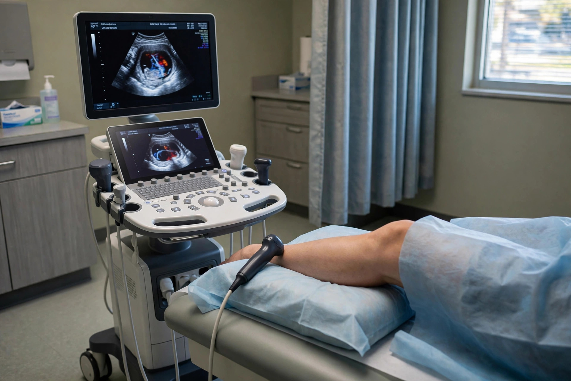
What it is
A leg Doppler (venous duplex) ultrasound uses sound waves to check the deep veins of the leg for a blood clot (deep vein thrombosis, DVT) and to assess vein valves in varicose vein problems.
Why it is done
It is the standard first test when a DVT is suspected — for example with a painful, swollen calf — and is also used to map varicose veins before treatment.
How to prepare
There is no preparation. Wear loose clothing or shorts so the leg can be examined from groin to ankle.
What happens during the procedure
Gel is applied and the probe is pressed along the leg at intervals; pressing the vein gently is part of the test, because a healthy vein squashes flat and a clot prevents this. It usually takes 20 to 30 minutes per leg and is painless apart from firm pressure.
Afterwards — recovery & results
You can leave straight away. Because suspected DVT is time-sensitive, the result is usually communicated the same day so treatment, if needed, can start promptly.
Risks & considerations
There are no known risks from the scan itself. Occasionally the result is uncertain — for example if the calf veins are hard to see — and a repeat scan a few days later may be arranged.
Related guides on MedicalVideos.com
Frequently asked questions
I had a blood test (D-dimer) as well — why both?
The blood test helps decide how likely a clot is. If it is raised, or the clinical suspicion is strong, the ultrasound is used to look for the clot directly.
Can the scan miss a clot?
It is very good for clots in the main deep veins above the knee. Small calf-vein clots can be harder to see, which is why a repeat scan is sometimes booked.
Related procedures — Imaging & Scans
Browse the L section of the A–Z list or return to the Procedures & Tests hub.