
- Physical Examination
- Surgical Examination
- Ophthalmology
- Clinical Skills
- Orthopedics
- Surgery Videos
- Laparoscopy
- Pediatrics
- Funny Videos
- Cardiothoracic Surgery
- Nursing Videos
- Plastic Surgery
- Otorhinolaryngology
- Histology and Histopathology
- Neurosurgery
- Dermatology
- Pediatric Surgery
- Urology
- Dentistry
- Oncology and Cancers
- Anatomy Videos
- Health and Fitness
- Radiology
- Anaesthesia
- Physical Therapy
- Pharmacology
- Interventional Radiology
- Cardiology
- Endocrinology
- Gynecology
- Emergency Medicine
- Psychiatry and Psychology
- Childbirth Videos
- General Medical Videos
- Nephrology
- Physiology
- Diet and Food Health
- Diabetes Mellitus
- Neurology
- Women Health
- Osteoporosis
- Gastroenterology
- Pulmonology
- Hematology
- Rheumatology
- Toxicology
- Nuclear Medicine
- Infectious Diseases
- Vascular Disease
- Reproductive Health
- Burns and Wound Healing
- Kidney Zone Podcast
- Other
MRI scan — spine
Category: Imaging & Scans · All imaging & scans
What it is
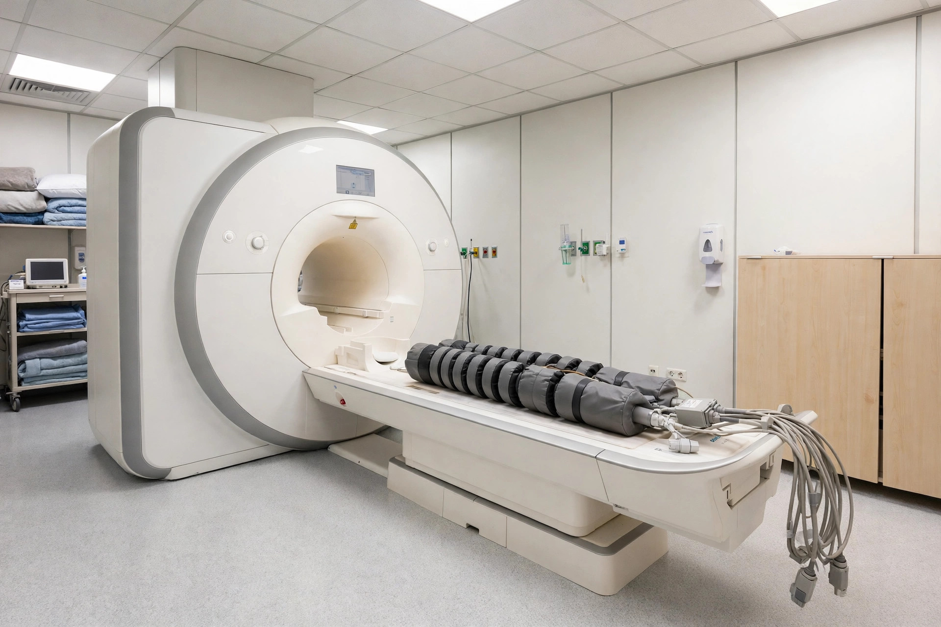
An MRI scan of the spine uses a magnet and radio waves to picture the spinal bones, discs, spinal cord and nerves in fine detail, without using X-rays.
Why it is done
It is commonly used for persistent back or neck pain with leg or arm symptoms, suspected slipped disc or trapped nerve, and to look for infection, tumours or spinal cord problems.
How to prepare
You will complete a metal-safety questionnaire and remove metal items. Most people do not need to fast. Tell the team if you have implants, a pacemaker or anxiety about enclosed spaces.
What happens during the procedure
You lie on your back on a table that moves into the scanner. The machine is noisy and you will wear ear protection. Keeping still is important; the scan usually takes 20 to 40 minutes depending on how much of the spine is scanned.
Afterwards — recovery & results
You can go home afterwards and carry on as normal. The report goes to your doctor, who will relate the pictures to your symptoms — many scan findings, such as disc bulges, are also seen in people with no pain at all.
Risks & considerations
MRI uses no ionising radiation. Risks are mainly those of the magnet for people with certain implants, and discomfort or anxiety during the scan. Contrast dye is sometimes used and is used cautiously in severe kidney disease.
Related guides on MedicalVideos.com
Frequently asked questions
My scan shows a disc bulge — does that mean I need surgery?
Not usually. Disc bulges are very common in people without any symptoms. Your doctor interprets the scan together with your examination and will discuss the options, which often start with non-surgical treatment.
How is MRI different from CT for the spine?
MRI shows discs, nerves and the spinal cord much more clearly, while CT shows bone detail well and is quicker. Your doctor chooses based on the question being asked.
Related procedures — Imaging & Scans
Browse the M section of the A–Z list or return to the Procedures & Tests hub.