
- Physical Examination
- Surgical Examination
- Ophthalmology
- Clinical Skills
- Orthopedics
- Surgery Videos
- Laparoscopy
- Pediatrics
- Funny Videos
- Cardiothoracic Surgery
- Nursing Videos
- Plastic Surgery
- Otorhinolaryngology
- Histology and Histopathology
- Neurosurgery
- Dermatology
- Pediatric Surgery
- Urology
- Dentistry
- Oncology and Cancers
- Anatomy Videos
- Health and Fitness
- Radiology
- Anaesthesia
- Physical Therapy
- Pharmacology
- Interventional Radiology
- Cardiology
- Endocrinology
- Gynecology
- Emergency Medicine
- Psychiatry and Psychology
- Childbirth Videos
- General Medical Videos
- Nephrology
- Physiology
- Diet and Food Health
- Diabetes Mellitus
- Neurology
- Women Health
- Osteoporosis
- Gastroenterology
- Pulmonology
- Hematology
- Rheumatology
- Toxicology
- Nuclear Medicine
- Infectious Diseases
- Vascular Disease
- Reproductive Health
- Burns and Wound Healing
- Kidney Zone Podcast
- Other
Pleural biopsy
Category: Lung & Breathing Tests · All lung & breathing tests
What it is
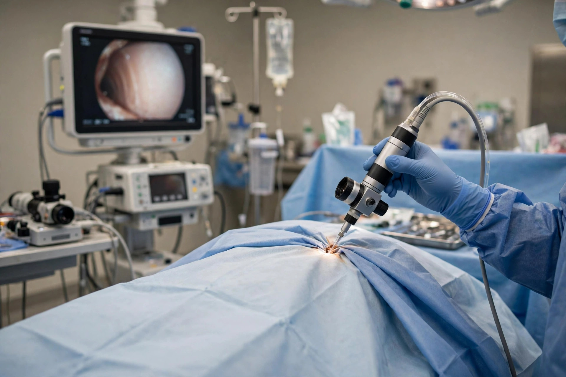
A pleural biopsy samples the lining of the chest (pleura) when fluid tests and scans have not explained a pleural effusion or pleural thickening. It can be done with a needle under imaging, or more usually today by medical thoracoscopy — a camera inserted through a small chest cut while you are sedated, allowing directed biopsies and drainage in the same session.
Why it is done
It is used mainly when cancer (including asbestos-related mesothelioma) or tuberculosis is suspected in the pleura and fluid cytology has been negative or inconclusive.
How to prepare
You will have CT imaging, blood tests and clotting checks. Medical thoracoscopy requires fasting and sedation; needle biopsy uses local anaesthetic alone. Tell the team about blood thinners, allergies and previous chest surgery or pleural procedures.
What happens during the procedure
For thoracoscopy: after sedation and local anaesthetic, a small cut admits the thoracoscope; the fluid is drained, the pleura inspected, and biopsies taken from abnormal areas. Talc may be puffed in to stick the lung to the chest wall (pleurodesis) if appropriate. A chest drain is left for a day or two. The session takes about 45 to 60 minutes.
Afterwards — recovery & results
You stay in hospital until the drain is removed and a chest X-ray is satisfactory. Samples take about one to two weeks (longer if TB cultures are needed). Your team will explain results and the treatment plan in clinic.
Risks & considerations
Risks include bleeding, infection, persistent air leak, fever after talc, and — rarely — spread of tumour along the biopsy tract (radiotherapy is sometimes offered to the site in mesothelioma). Serious complications are uncommon in experienced centres.
Related guides on MedicalVideos.com
Frequently asked questions
Why wasn't the fluid test enough?
Cancer and TB cells do not always shed into the fluid. Biopsying the lining itself finds the diagnosis in most cases where fluid testing cannot.
What is pleurodesis and why combine it?
If fluid keeps re-accumulating, talc inflames the pleural surfaces so they stick together and leave no space for fluid. Doing it during the same procedure saves a second intervention.
Related procedures — Lung & Breathing Tests
Browse the P section of the A–Z list or return to the Procedures & Tests hub.