
- Physical Examination
- Surgical Examination
- Ophthalmology
- Clinical Skills
- Orthopedics
- Surgery Videos
- Laparoscopy
- Pediatrics
- Funny Videos
- Cardiothoracic Surgery
- Nursing Videos
- Plastic Surgery
- Otorhinolaryngology
- Histology and Histopathology
- Neurosurgery
- Dermatology
- Pediatric Surgery
- Urology
- Dentistry
- Oncology and Cancers
- Anatomy Videos
- Health and Fitness
- Radiology
- Anaesthesia
- Physical Therapy
- Pharmacology
- Interventional Radiology
- Cardiology
- Endocrinology
- Gynecology
- Emergency Medicine
- Psychiatry and Psychology
- Childbirth Videos
- General Medical Videos
- Nephrology
- Physiology
- Diet and Food Health
- Diabetes Mellitus
- Neurology
- Women Health
- Osteoporosis
- Gastroenterology
- Pulmonology
- Hematology
- Rheumatology
- Toxicology
- Nuclear Medicine
- Infectious Diseases
- Vascular Disease
- Reproductive Health
- Burns and Wound Healing
- Kidney Zone Podcast
- Other
Thyroid ultrasound
Category: Imaging & Scans · All imaging & scans
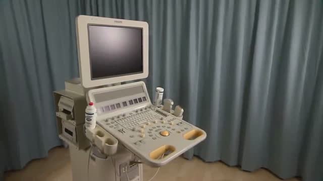
What it is
A thyroid ultrasound uses sound waves to picture the thyroid gland in the front of the neck, showing its size and any lumps (nodules) within it.
Why it is done
It is used to investigate a lump or swelling in the neck, an enlarged thyroid, or abnormal thyroid blood tests, and to guide a fine-needle sample of a nodule.
How to prepare
No preparation is needed. You may be asked to remove necklaces and wear a gown or open collar.
What happens during the procedure
You lie back with your neck slightly extended, gel is applied and the probe is moved over the front of the neck. It is painless and takes about 15 to 20 minutes.
Afterwards — recovery & results
You can go home immediately. The report goes to your doctor, who will combine it with your blood tests and examination to decide whether a needle sample or follow-up scan is needed.
Risks & considerations
There are no known risks from the scan. Many thyroid nodules found on ultrasound are harmless, so finding one does not automatically mean treatment is required.
Watch: Thyroid ultrasound videos
Real procedure videos from the MedicalVideos.com library. Some clinical footage is graphic — that is the nature of a medical video library.
Related guides on MedicalVideos.com
Frequently asked questions
The scan found a nodule — should I worry?
Thyroid nodules are very common and most are benign. Size and appearance guide whether a needle sample is recommended.
Will I need a biopsy?
Only if the nodule's appearance or size meets the criteria the specialist uses. Many nodules simply need a follow-up scan.
Related procedures — Imaging & Scans
Browse the T section of the A–Z list or return to the Procedures & Tests hub.