
- Physical Examination
- Surgical Examination
- Ophthalmology
- Clinical Skills
- Orthopedics
- Surgery Videos
- Laparoscopy
- Pediatrics
- Funny Videos
- Cardiothoracic Surgery
- Nursing Videos
- Plastic Surgery
- Otorhinolaryngology
- Histology and Histopathology
- Neurosurgery
- Dermatology
- Pediatric Surgery
- Urology
- Dentistry
- Oncology and Cancers
- Anatomy Videos
- Health and Fitness
- Radiology
- Anaesthesia
- Physical Therapy
- Pharmacology
- Interventional Radiology
- Cardiology
- Endocrinology
- Gynecology
- Emergency Medicine
- Psychiatry and Psychology
- Childbirth Videos
- General Medical Videos
- Nephrology
- Physiology
- Diet and Food Health
- Diabetes Mellitus
- Neurology
- Women Health
- Osteoporosis
- Gastroenterology
- Pulmonology
- Hematology
- Rheumatology
- Toxicology
- Nuclear Medicine
- Infectious Diseases
- Vascular Disease
- Reproductive Health
- Burns and Wound Healing
- Kidney Zone Podcast
- Other













Da Vinci Robotic Hysterectomy

Educational only: videos on MedicalVideos.com are for general information and are not medical advice. Always consult your own doctor about your health.
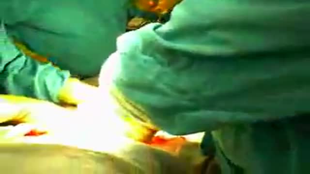
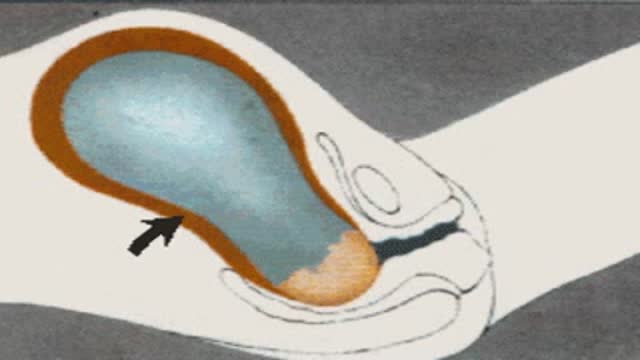
The surgical video details a robotic assisted hysterectomy in a patient with early stage endometrial/uterine cancer. Anatomy of the pelvis and the technique of a robotic hysterectomy is demonstrated in this video.
This video in 30 seconds
This surgical video details a robotic assisted hysterectomy in a patient with early stage endometrial or uterine cancer. It demonstrates the anatomy of the pelvis and the technique of a robotic hysterectomy.
Key points
- Robotic assisted hysterectomy is detailed
- The patient has early stage endometrial or uterine cancer
- Anatomy of the pelvis is demonstrated
- The technique of robotic hysterectomy is demonstrated
Video Transcript
A narrated robotic-assisted hysterectomy using the da Vinci system in a patient with early-stage uterine cancer. The surgeon describes docking the robot and works through the pelvic anatomy and dissection step by step.
Read the full transcript
DaVinci robotic hysterectomy, surgeon M. Patrick Lowe. After successful insertion of the laparoscopic trochars and docking of the robot, the procedure is ready to begin. For this procedure, we have chosen to use hot shears with monopolar cottery and a Maryland dissector with bipolar cottery. The initial step in the procedure is to open the peritoneum overlying the avascular space of grays on the right pelvic sidewall. This allows for identification of the iliac vessels as well as for cauterization and division of the round ligament. Next, the avascular space of grays is divided to the bifurcation of the iliac vessels using the endoshears with monopolar cottery.
The infundibulopelvic ligament is grasped, elevated, and skeletonized using monopolar cottery with the endoshears. The skeletonized ovarian artery and vein are now sealed with the ligature atlas vessel sealing device. The posterior leaf of the broad ligament is then divided to the posterior surface of the uterus using general dissection and monopolar cottery with the endoshears. The round ligament is cauterized with bipolar cottery and subsequently divided with hot shears and monopolar cottery. Now the anterior leaf of the broad ligament is divided allowing for development of the bladder flap. This is accomplished using monopolar cottery in the tips of endoshears to develop the space using general sweeping motions of the robotic arm, allowing for delicate controlled dissection in this area.
The left round ligament is cauterized with bipolar cottery and subsequently divided using the endoshears and monopolar cottery. The retroperitoneal space is then developed to the level of the bifurcation of the aliac vessels as shown here. A window is created in the medial leaf of the broad ligament using hot shears and monopolar cottery with further development via blunt dissection. The skeletonized ovarian artery and vein are now sealed with the ligature atlas vessel sealing device. The posterior leaf of the broad ligament is then incised to the posterior surface of the uterus.
The left uterine artery is skeletonized using blunt and sharp dissection with monopolar cottery and hot shears. The left uterine artery is now sealed with the ligature atlas vessel sealing system. Additional dissection is performed along the cardinal uterus acro complex utilizing hot shears and monopolar cottery. The right uterine artery is now sealed with the ligature atlas vessel sealing system. The ring of the uterine manipulator is now visualized in an incision between the uterus acro ligaments when the posterior vaginal wall is performed. The outline of the cervical cup is followed in a circumferential fashion and sizing the intervening vaginal tissue.
The uterus is then removed via the vagina through general traction on the uterine manipulator. The vaginal cuff is enclosed in a running continuous fashion utilizing a zero vicaral suture in a CT1 needle with the cuff angle secured with a laparoti. We use the suture cut needle driver and either a Maryland bipolar grasper or a second mega needle driver to close the vaginal cuff. Here a second laparoti is placed at the opposite vaginal cuff angle securing the suture. The patient was discharged home from the hospital the day after surgery without postoperative complications.
Transcript auto-generated from the video’s audio for accessibility and study. Educational content only — not medical advice.
Video Overview
From our Gynecology collection: Pelvic exams, Pap smears, IUD insertion, hysteroscopy and women's health procedures explained step by step. Browse all Gynecology videos →
Medical Videos
Free medical and surgery videos for patients, students and professionals — trusted since 2007.
Founded by Dr. Mohamed Ibrahim, MD, PhD — physician-scientist.
Follow us on Facebook · Instagram
SORT BY-
Top Comments
-
Latest comments