
- Procedures & Tests
- Symptoms A-Z
- Symptom Checker
- Kidney Diet
- First Aid
- Anatomy Atlas
- Ask MedicalVideos
- Drug Guide
- Lab Values
- Vital Signs
- Muscles A–Z
- Clinical Signs
- Medical Abbreviations
- Medical Terminology
- Physical Examination
- Surgical Examination
- Ophthalmology
- Clinical Skills
- Orthopedics
- Surgery Videos
- Laparoscopy
- Pediatrics
- Medical Humor
- Cardiothoracic Surgery
- Nursing Videos
- Plastic Surgery
- Otorhinolaryngology
- Histology and Histopathology
- Neurosurgery
- Dermatology
- Pediatric Surgery
- Urology
- Dentistry
- Oncology and Cancers
- Anatomy Videos
- Health and Fitness
- Radiology
- Anaesthesia
- Physical Therapy
- Pharmacology
- Interventional Radiology
- Cardiology
- Endocrinology
- Gynecology
- Emergency Medicine
- Psychiatry and Psychology
- Childbirth Videos
- General Medical Videos
- Nephrology
- Physiology
- Diet and Food Health
- Diabetes Mellitus
- Neurology
- Women Health
- Osteoporosis
- Gastroenterology
- Pulmonology
- Hematology
- Rheumatology
- Toxicology
- Nuclear Medicine
- Infectious Diseases
- Vascular Disease
- Reproductive Health
- Burns and Wound Healing
- Kidney Zone Podcast
- Other





Ultrasound-guided internal jugular cannulation
kareem S. Ultrasound-guided internal jugular cannulation [video]. MedicalVideos.com; 2024 Jan 17 [cited 2026 Nov 21]. Available from: https://www.medicalvideos.com/watch/ultrasound-guided-internal-jugular-cannulation_8f1355681e814c317679.html
kareem, S. (2024, January 17). Ultrasound-guided internal jugular cannulation [Video]. MedicalVideos.com. https://www.medicalvideos.com/watch/ultrasound-guided-internal-jugular-cannulation_8f1355681e814c317679.html
kareem, samer. “Ultrasound-guided internal jugular cannulation.” MedicalVideos.com, 17 Jan. 2024, https://www.medicalvideos.com/watch/ultrasound-guided-internal-jugular-cannulation_8f1355681e814c317679.html. Accessed 21 Nov. 2026.
@misc{medicalvideos2024_8f1355681e814c317679,
  author = {kareem, samer},
  title = {Ultrasound-guided internal jugular cannulation},
  publisher = {MedicalVideos.com},
  year = {2024},
  month = {1},
  url = {https://www.medicalvideos.com/watch/ultrasound-guided-internal-jugular-cannulation_8f1355681e814c317679.html},
  note = {Video},
  urldate = {2026-11-21}
}Vancouver (NLM) is the manuscript standard for medical journals. RIS imports into EndNote, Zotero and Mendeley.

Educational only: videos on MedicalVideos.com are for general information and are not medical advice. Always consult your own doctor about your health.
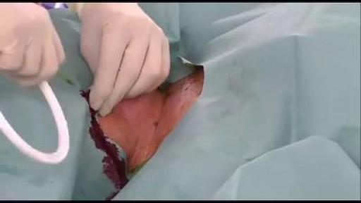
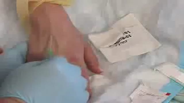
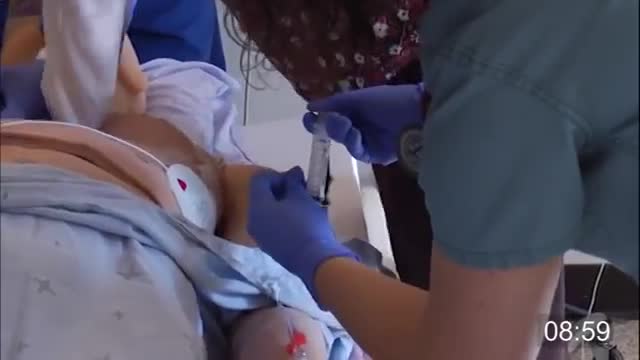
This narrated demonstration shows ultrasound-guided cannulation of the internal jugular vein. The vein is identified beside the carotid artery in short and long axis views, a sterile field and covered transducer are prepared, and the needle is advanced in-plane so its whole length stays visible. Venous return confirms placement before the Seldinger steps finish the line.
Video Transcript
This narrated demonstration shows ultrasound-guided cannulation of the internal jugular vein. It covers identifying the vein beside the carotid artery on short and long axis views, preparing a sterile field and covered transducer, inserting the needle in-plane so its whole length is seen, confirming venous blood return, and completing the catheter insertion by the Seldinger technique.
Read the full transcript
Before proceeding with the vascular puncture, it is appropriate to perform an ultrasound exam of the cervical region.
On the short or transversal axis, the internal jugular vein is identified as a vessel antirelateral to the common carotid artery, which is compressible and non-pulsatile.
On the long or longitudinal axis, the internal jugular is more superficial than the common carotid. To ensure safety during the puncture, it is convenient to move the transducer laterally, until the carotid can no longer be seen under the jugular. After removing the remnants of gel, the skin is prepared with an antiseptic solution. The cannulator prepares the material and surgical field using an aseptic technique. The ultrasound transducer is then covered using a sterile sheath to avoid contaminating the surgical field. Using sterile saline solution as a coupling medium, the internal jugular vein is relocated on the long axis.
To minimize the risks arising from possible posterior vein wall puncture, it is better to avoid the planes in which the carotid appears underneath. The celled-inja needle is inserted in-plane to observe the entire length of the needle. After verifying the intravascular location of the needle tip, proceed to aspirate with the syringe and confirm that venous blood reflux is observed.
The transducer is then removed and the guide wire introduced through the needle. Confirming that the guide wire is in an intravascular position in the internal jugular vein is recommended.
Once the guide wire is inserted, continue with the celled-inja technique for catheter insertion.
Transcript auto-generated from the video’s audio for accessibility and study. Educational content only — not medical advice.
Related reading
Save videos for later
Create a free account to build your own watch-later list and playlists, and pick up where you left off on any device.
Medical Videos
Free medical and surgery videos for patients, students and professionals — trusted since 2007.
Founded by Dr. Mohamed Ibrahim, MD, PhD — physician-scientist.
Follow us on Facebook · Instagram
SORT BY-
Top Comments
-
Latest comments