
- Physical Examination
- Surgical Examination
- Ophthalmology
- Clinical Skills
- Orthopedics
- Surgery Videos
- Laparoscopy
- Pediatrics
- Funny Videos
- Cardiothoracic Surgery
- Nursing Videos
- Plastic Surgery
- Otorhinolaryngology
- Histology and Histopathology
- Neurosurgery
- Dermatology
- Pediatric Surgery
- Urology
- Dentistry
- Oncology and Cancers
- Anatomy Videos
- Health and Fitness
- Radiology
- Anaesthesia
- Physical Therapy
- Pharmacology
- Interventional Radiology
- Cardiology
- Endocrinology
- Gynecology
- Emergency Medicine
- Psychiatry and Psychology
- Childbirth Videos
- General Medical Videos
- Nephrology
- Physiology
- Diet and Food Health
- Diabetes Mellitus
- Neurology
- Women Health
- Osteoporosis
- Gastroenterology
- Pulmonology
- Hematology
- Rheumatology
- Toxicology
- Nuclear Medicine
- Infectious Diseases
- Vascular Disease
- Reproductive Health
- Burns and Wound Healing
- Kidney Zone Podcast
- Other


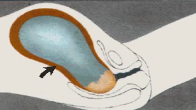
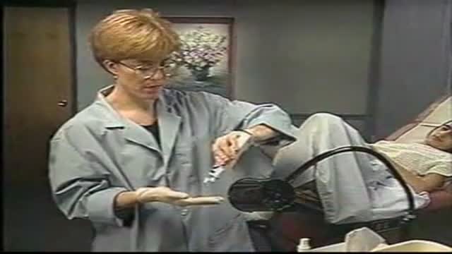
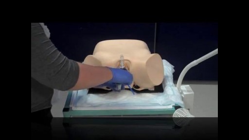
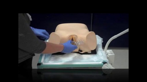
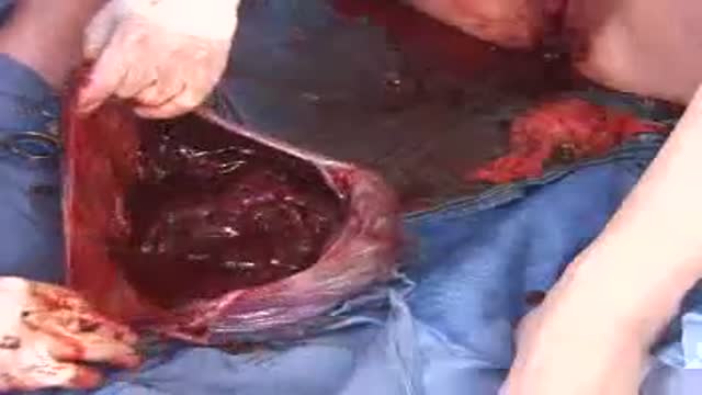
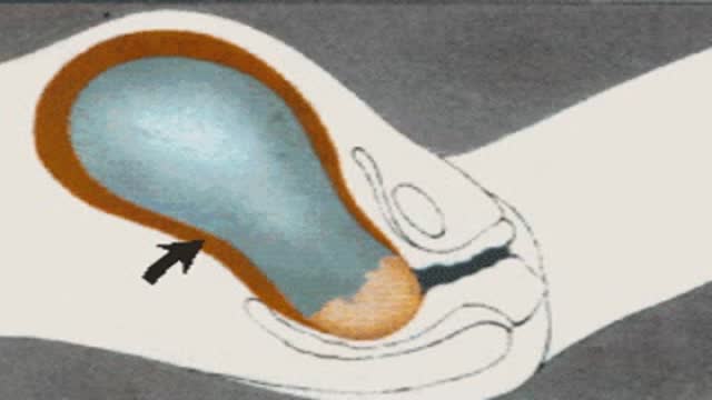
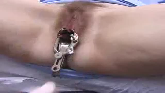
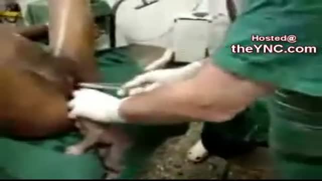




Vaginal Hysterectomy Procedure

Educational only: videos on MedicalVideos.com are for general information and are not medical advice. Always consult your own doctor about your health.
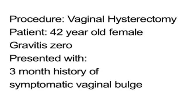
Vaginal Hysterectomy Procedure of a 42 years old female patient with a 3 months history of symptomatic vaginal bulge
Video Transcript
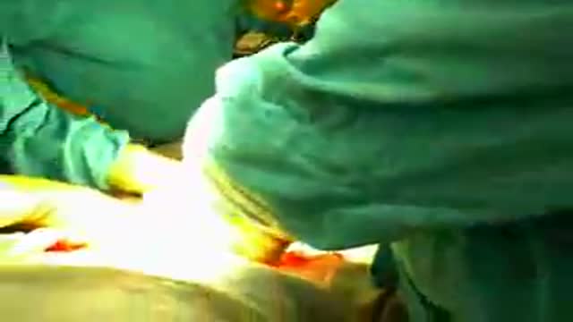
A narrated vaginal hysterectomy performed on a 42-year-old woman with a symptomatic vaginal bulge. The surgeon talks through the posterior colpotomy, vessel sealing, and the stepwise removal of the uterus.
Read the full transcript
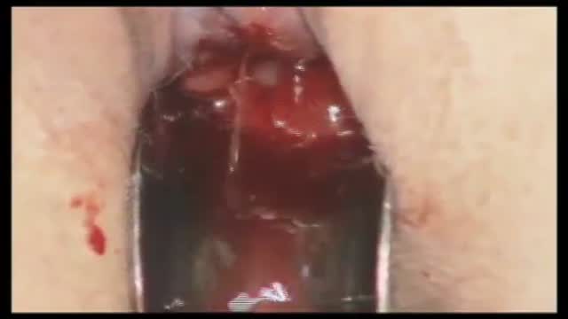
This procedure is a vaginal hysterectomy utilizing the plasma seal open forceps. This is a case of a 42-year-old white female, Gravita-0, with a three-month history of a symptomatic vaginal bulge. As we start the vaginal hysterectomy, a leji clamp is used to co-opt the anterior and posterior cervix. Quarter percent marcane, approximately 5 to 10 cc's, is injected in the posterior fornix for mucosal hemostasis prior to the posterior copotomy. This is with epinephrine. Once the posterior copotomy is made, a running interlocking suture of Xerovigral is used for hemostasis of the posterior vaginal wall.
This is done in my practice due to the extensive repairs that are required after the hysterectomy and the length of the possible bleeding that could occur during these repairs. The blade of the Bretsky-Navichil retractor is in place in the posterior cul-de-sac. The use of the plasma seal open forceps begins. The Bretsky-Navichil retractors are of great help during the surgical procedure to apply retraction to the lateral sidewall and the posterior vaginal wall for surgeon visualization.
As we do the left uter-sacral ligament complex, the plasma seal open forceps are initially used on coagulation and then the foot pedal is used on the cutting current. On the patient's right side, the same uter-sacral ligament pedicle is grasped and coagged and cut. Additional use of the plasma seal open forceps is seen here due to the large size of this pedicle. Weighted speculum is then used instead of the Bretsky-Navichil retractor in the posterior fornix. The cardinal ligaments are then grasped with the plasma seal forceps and coagulated and cut.
The same cardinal uter-sacral ligament is grasped with the forceps on the patient's right side. Serial coagulation and cutting is performed along the cervical vaginal junction on the right and left side. A longer-nosed weighted speculum is then positioned in the posterior vaginal wall and into the posterior copotomy incision. The serial clamping is performed until the cervical vaginal junction at the recess of the bladder is reached. Bovic coagulation and cutting is then used to create the vesicovaginal space. During this surgical procedure, the cervix was not circumscribed with the scalpel as seen with many vaginal hysterectomies.
The anterior copotomy is made without difficulty. The Diva retractor is placed with the anterior copotomy to protect the bladder and the peritoneum is co-apted both anteriorly and posteriorly and the bladder pillars all into the forceps. This is performed bilaterally with excellent hemostasis. The plasma seal open forceps theoretically save time because we do not have to put on a clamp, cut the pedicle, and then tie the pedicle with a suture. Procedural efficiency is important in the operating room for the patient should have the least invasive surgery in the least amount of time that is safe and effective as well as decreasing exposure to anesthesia.
With this new technology demonstrated today, I would hope that if you are in a training institution that your residents still will learn traditional surgical removal of the uterus with clamping, cutting, and tying, but as they advance in their training, this type of procedure is ideal for patient care. After delivering most of the uterus, the tubular ovarian ligaments are then approached, and it was my choice during this procedure to clamp these pedicles with the traditional hainy clamp with suture ligature. Due to this patient having a rather large uterus with significant blood flow to the uterus, I felt more comfortable placing this suture due to the possibility of the vessels being larger than 7 millimeters.
The hainy clamp was also used in the right tubular ovarian ligament. The hysterectomy is now complete. This large irregular uterus was safely removed with the plasma seal open forceps in approximately 36 minutes.
Transcript auto-generated from the video’s audio for accessibility and study. Educational content only — not medical advice.
Video Overview
From our Gynecology collection: Pelvic exams, Pap smears, IUD insertion, hysteroscopy and women's health procedures explained step by step. Browse all Gynecology videos →
Medical Videos
Free medical and surgery videos for patients, students and professionals — trusted since 2007.
Founded by Dr. Mohamed Ibrahim, MD, PhD — physician-scientist.
Follow us on Facebook · Instagram
SORT BY-
Top Comments
-
Latest comments