- Physical Examination
- Surgical Examination
- Ophthalmology
- Clinical Skills
- Orthopedics
- Surgery Videos
- Laparoscopy
- Pediatrics
- Funny Videos
- Cardiothoracic Surgery
- Nursing Videos
- Plastic Surgery
- Otorhinolaryngology
- Histology and Histopathology
- Neurosurgery
- Dermatology
- Pediatric Surgery
- Urology
- Dentistry
- Oncology and Cancers
- Anatomy Videos
- Health and Fitness
- Radiology
- Anaesthesia
- Physical Therapy
- Pharmacology
- Interventional Radiology
- Cardiology
- Endocrinology
- Gynecology
- Emergency Medicine
- Psychiatry and Psychology
- Childbirth Videos
- General Medical Videos
- Nephrology
- Physiology
- Diet and Food Health
- Diabetes Mellitus
- Neurology
- Women Health
- Osteoporosis
- Gastroenterology
- Pulmonology
- Hematology
- Rheumatology
- Toxicology
- Nuclear Medicine
- Infectious Diseases
- Vascular Disease
- Reproductive Health
- Burns and Wound Healing
- Kidney Zone Podcast
- Other
Calcaneus
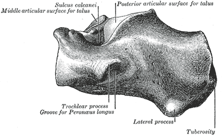
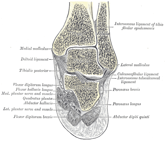
Gray's Anatomy (1918) plate, public domain
The calcaneus is the heel bone — the largest tarsal and the first part of the foot to strike the ground when walking. It sits below the talus, projecting backwards to form the heel, where the Achilles tendon attaches. Its upper surface carries the joints of the subtalar joint, which lets the foot tilt side to side on uneven ground.
Key facts
- The calcaneus is the largest bone of the foot.
- It forms the heel and takes the Achilles tendon.
- It supports the talus above it at the subtalar joint.
- A shelf on its inner side (sustentaculum tali) supports the talus and guides tendons.
- The plantar fascia attaches to its underside.
What can go wrong
Calcaneal fractures are classic injuries of falling from a height onto the heels — and the other heel and the spine are checked for matching injuries. Heel pain itself is far more often from the plantar fascia's attachment (plantar fasciitis) or from irritation where the Achilles tendon joins, than from the bone.
Related procedures & tests
Related symptoms
Related muscles
Frequently asked questions
Why check the spine after a heel fracture?
The force that breaks a heel in a fall travels straight up the leg; the same impact can compress vertebrae in the lower back, so both heels and the spine are examined and imaged.
What is a heel spur?
A small bony outgrowth under the calcaneus where the plantar fascia attaches. Spurs are common and are usually a sign of long-standing pull at the attachment rather than the direct cause of pain.
Educational information only. This atlas explains normal anatomy and describes conditions in general terms; it is not medical advice and does not replace assessment by a qualified clinician. If you have symptoms that worry you, seek medical care.