- Physical Examination
- Surgical Examination
- Ophthalmology
- Clinical Skills
- Orthopedics
- Surgery Videos
- Laparoscopy
- Pediatrics
- Funny Videos
- Cardiothoracic Surgery
- Nursing Videos
- Plastic Surgery
- Otorhinolaryngology
- Histology and Histopathology
- Neurosurgery
- Dermatology
- Pediatric Surgery
- Urology
- Dentistry
- Oncology and Cancers
- Anatomy Videos
- Health and Fitness
- Radiology
- Anaesthesia
- Physical Therapy
- Pharmacology
- Interventional Radiology
- Cardiology
- Endocrinology
- Gynecology
- Emergency Medicine
- Psychiatry and Psychology
- Childbirth Videos
- General Medical Videos
- Nephrology
- Physiology
- Diet and Food Health
- Diabetes Mellitus
- Neurology
- Women Health
- Osteoporosis
- Gastroenterology
- Pulmonology
- Hematology
- Rheumatology
- Toxicology
- Nuclear Medicine
- Infectious Diseases
- Vascular Disease
- Reproductive Health
- Burns and Wound Healing
- Kidney Zone Podcast
- Other
Fibula
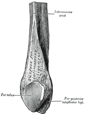
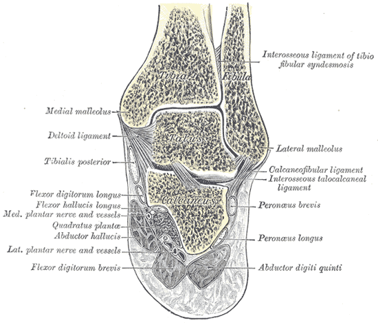
Gray's Anatomy (1918) plate, public domain
The fibula is the slender bone on the outer side of the lower leg. It carries little weight — its roles are to anchor the ligaments of the knee and ankle, to form the outer side of the ankle joint, and to give origin to muscles of the leg and foot. Its head at the knee is a landmark, with the common fibular nerve winding around it.
Key facts
- The fibula bears only a small fraction of the leg's load.
- Its lower end forms the outer ankle bone (lateral malleolus).
- Its head gives attachment to the biceps femoris and the knee's outer ligament.
- The common fibular (peroneal) nerve winds around its neck — a vulnerable spot.
- Sections of fibula can be borrowed as grafts for reconstructing other bones.
What can go wrong
The fibula most often breaks at the ankle as part of ankle fractures and sprains. A blow or prolonged pressure over its neck — from crossing legs, a tight cast or squatting — can bruise the nerve there and cause foot drop, which usually recovers. Isolated shaft fractures from direct blows are also seen.
Related procedures & tests
Related symptoms
Related muscles
Watch: related videos
Frequently asked questions
If the fibula carries so little weight, why keep it?
The ankle needs its outer wall: without the fibula's lower end the ankle joint is unstable. It also serves as a strut and muscle anchor along the whole leg.
Why does a knock below the knee sometimes cause foot drop?
The common fibular nerve wraps around the fibula's neck just under the skin. Bruising it interrupts the signal to the muscles that lift the foot, producing a temporary foot drop.
Educational information only. This atlas explains normal anatomy and describes conditions in general terms; it is not medical advice and does not replace assessment by a qualified clinician. If you have symptoms that worry you, seek medical care.