- Physical Examination
- Surgical Examination
- Ophthalmology
- Clinical Skills
- Orthopedics
- Surgery Videos
- Laparoscopy
- Pediatrics
- Funny Videos
- Cardiothoracic Surgery
- Nursing Videos
- Plastic Surgery
- Otorhinolaryngology
- Histology and Histopathology
- Neurosurgery
- Dermatology
- Pediatric Surgery
- Urology
- Dentistry
- Oncology and Cancers
- Anatomy Videos
- Health and Fitness
- Radiology
- Anaesthesia
- Physical Therapy
- Pharmacology
- Interventional Radiology
- Cardiology
- Endocrinology
- Gynecology
- Emergency Medicine
- Psychiatry and Psychology
- Childbirth Videos
- General Medical Videos
- Nephrology
- Physiology
- Diet and Food Health
- Diabetes Mellitus
- Neurology
- Women Health
- Osteoporosis
- Gastroenterology
- Pulmonology
- Hematology
- Rheumatology
- Toxicology
- Nuclear Medicine
- Infectious Diseases
- Vascular Disease
- Reproductive Health
- Burns and Wound Healing
- Kidney Zone Podcast
- Other
Cerebral Ventricles
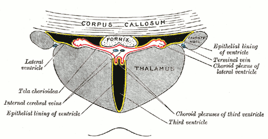
Gray's Anatomy (1918) plate, public domain
The cerebral ventricles are the interconnected fluid-filled cavities within the brain: two large C-shaped lateral ventricles in the hemispheres, the midline third ventricle, and the fourth ventricle in the brainstem, continuous with the spinal cord's central canal. They hold cerebrospinal fluid (CSF), made by the choroid plexuses inside them, which cushions the brain, removes waste and maintains the chemical environment. CSF flows through narrow channels between the ventricles and out around the brain and cord.
Key facts
- The brain makes about 500 mL of CSF a day, though only about 150 mL is present at any time — it is constantly renewed.
- CSF circulation has a direction: lateral ventricles → third → aqueduct → fourth → around the brain and cord, then into the veins.
- The narrow cerebral aqueduct is the system's most blockage-prone point.
- Floating in CSF, the brain's effective weight drops from about 1,400 g to roughly 50 g, reducing pressure on its base.
What can go wrong
Blocked flow or absorption enlarges the ventricles (hydrocephalus): in babies the head expands, while in adults raised pressure causes headache, vomiting and visual problems; a chronic form in older adults (normal pressure hydrocephalus) gives a triad of walking difficulty, urinary incontinence and cognitive slowing, and can improve with a shunt. Bleeding or infection can block the pathways, and tumours can obstruct the narrow channels.
Related conditions
Related procedures & tests
Related symptoms
Frequently asked questions
What is a shunt for hydrocephalus?
A fine tube with a valve, usually from a ventricle to the abdomen, that drains excess CSF and relieves pressure when the natural pathways are blocked.
Why is CSF sampled by lumbar puncture?
Because it bathes the brain and cord, CSF reveals infections, inflammation, bleeding and pressure problems affecting the nervous system.
Educational information only. This atlas explains normal anatomy and describes conditions in general terms; it is not medical advice and does not replace assessment by a qualified clinician. If you have symptoms that worry you, seek medical care.