- Physical Examination
- Surgical Examination
- Ophthalmology
- Clinical Skills
- Orthopedics
- Surgery Videos
- Laparoscopy
- Pediatrics
- Funny Videos
- Cardiothoracic Surgery
- Nursing Videos
- Plastic Surgery
- Otorhinolaryngology
- Histology and Histopathology
- Neurosurgery
- Dermatology
- Pediatric Surgery
- Urology
- Dentistry
- Oncology and Cancers
- Anatomy Videos
- Health and Fitness
- Radiology
- Anaesthesia
- Physical Therapy
- Pharmacology
- Interventional Radiology
- Cardiology
- Endocrinology
- Gynecology
- Emergency Medicine
- Psychiatry and Psychology
- Childbirth Videos
- General Medical Videos
- Nephrology
- Physiology
- Diet and Food Health
- Diabetes Mellitus
- Neurology
- Women Health
- Osteoporosis
- Gastroenterology
- Pulmonology
- Hematology
- Rheumatology
- Toxicology
- Nuclear Medicine
- Infectious Diseases
- Vascular Disease
- Reproductive Health
- Burns and Wound Healing
- Kidney Zone Podcast
- Other
First Cuneiform
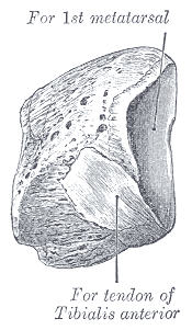
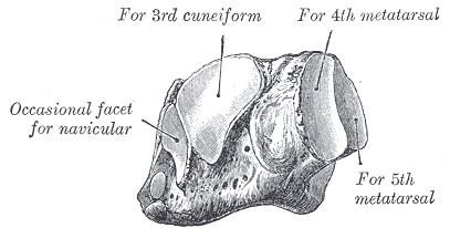
Gray's Anatomy (1918) plate, public domain
The first (medial) cuneiform is the largest of the three cuneiform bones of the midfoot. It sits between the navicular and the base of the big toe's metatarsal, forming the inner column of the foot that delivers push-off force when walking. The tibialis anterior tendon attaches partly to it.
Key facts
- It is the largest of the three cuneiforms.
- It forms the midfoot joint of the big-toe ray.
- It takes attachments from tibialis anterior and fibularis longus tendons.
- It is a key part of the Lisfranc joint line.
- Its strength underpins the medial arch's front pillar.
What can go wrong
The first cuneiform is involved in Lisfranc injuries of the midfoot, where the metatarsal bases shift on the cuneiforms after a twist or crush — injuries that need prompt recognition to protect the arch. Isolated fractures are uncommon.
Related procedures & tests
Related symptoms
Related muscles
Frequently asked questions
What is a Lisfranc injury?
Damage to the joints and ligaments between the cuneiforms/cuboid and the metatarsal bases — named after a Napoleonic-era surgeon. The second metatarsal base is the usual keystone that fails.
Why does the big-toe ray need such a solid midfoot bone?
Push-off at the end of each step funnels through the big toe; the medial cuneiform is the fixed base that lets the first metatarsal act as a lever.
Educational information only. This atlas explains normal anatomy and describes conditions in general terms; it is not medical advice and does not replace assessment by a qualified clinician. If you have symptoms that worry you, seek medical care.