- Physical Examination
- Surgical Examination
- Ophthalmology
- Clinical Skills
- Orthopedics
- Surgery Videos
- Laparoscopy
- Pediatrics
- Funny Videos
- Cardiothoracic Surgery
- Nursing Videos
- Plastic Surgery
- Otorhinolaryngology
- Histology and Histopathology
- Neurosurgery
- Dermatology
- Pediatric Surgery
- Urology
- Dentistry
- Oncology and Cancers
- Anatomy Videos
- Health and Fitness
- Radiology
- Anaesthesia
- Physical Therapy
- Pharmacology
- Interventional Radiology
- Cardiology
- Endocrinology
- Gynecology
- Emergency Medicine
- Psychiatry and Psychology
- Childbirth Videos
- General Medical Videos
- Nephrology
- Physiology
- Diet and Food Health
- Diabetes Mellitus
- Neurology
- Women Health
- Osteoporosis
- Gastroenterology
- Pulmonology
- Hematology
- Rheumatology
- Toxicology
- Nuclear Medicine
- Infectious Diseases
- Vascular Disease
- Reproductive Health
- Burns and Wound Healing
- Kidney Zone Podcast
- Other
First Metatarsal
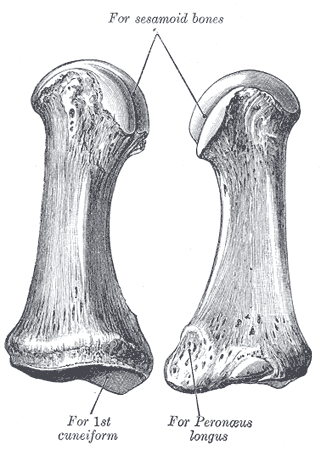
Gray's Anatomy (1918) plate, public domain
The first metatarsal is the big toe's forefoot bone — the shortest and by far the thickest metatarsal, built to take the final push of every step. Two small sesamoid bones sit under its head, cushioning it and guiding the big toe's flexor tendon. Its joint with the toe is the one affected by bunions and by hallux rigidus (stiff big toe).
Key facts
- It is the strongest metatarsal, carrying about 40% of forefoot load at push-off.
- Two sesamoids sit beneath its head in the flexor tendons.
- Its base forms a strong joint with the medial cuneiform.
- Bunions are a deformity at its head joint (the first MTP joint).
- The big toe's alignment determines efficient walking.
What can go wrong
When the first metatarsal angles away from the second, the big toe drifts towards the others — a bunion (hallux valgus) — which can become painful and make shoes difficult. Arthritis of its toe joint (hallux rigidus) stiffens push-off. Fractures are usually from direct blows or stubbing injuries.
Related procedures & tests
Related symptoms
Related muscles
Frequently asked questions
What is a bunion, in bone terms?
The first metatarsal tilts inwards while the big toe tilts outwards at their joint, so the metatarsal head becomes the prominent bump on the inner side of the foot.
Why does the big toe matter so much for walking?
It is the last part of the foot to leave the ground and acts as the lever for push-off; a stiff or painful big-toe joint changes the whole gait.
Educational information only. This atlas explains normal anatomy and describes conditions in general terms; it is not medical advice and does not replace assessment by a qualified clinician. If you have symptoms that worry you, seek medical care.