- Physical Examination
- Surgical Examination
- Ophthalmology
- Clinical Skills
- Orthopedics
- Surgery Videos
- Laparoscopy
- Pediatrics
- Funny Videos
- Cardiothoracic Surgery
- Nursing Videos
- Plastic Surgery
- Otorhinolaryngology
- Histology and Histopathology
- Neurosurgery
- Dermatology
- Pediatric Surgery
- Urology
- Dentistry
- Oncology and Cancers
- Anatomy Videos
- Health and Fitness
- Radiology
- Anaesthesia
- Physical Therapy
- Pharmacology
- Interventional Radiology
- Cardiology
- Endocrinology
- Gynecology
- Emergency Medicine
- Psychiatry and Psychology
- Childbirth Videos
- General Medical Videos
- Nephrology
- Physiology
- Diet and Food Health
- Diabetes Mellitus
- Neurology
- Women Health
- Osteoporosis
- Gastroenterology
- Pulmonology
- Hematology
- Rheumatology
- Toxicology
- Nuclear Medicine
- Infectious Diseases
- Vascular Disease
- Reproductive Health
- Burns and Wound Healing
- Kidney Zone Podcast
- Other
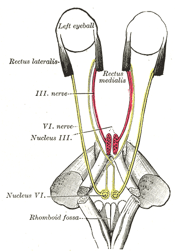
Oculomotor Nerve
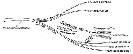
Gray's Anatomy (1918) plate, public domain
The oculomotor nerve (cranial nerve III) is the main nerve of eye movement. It supplies most of the muscles that move the eyeball — looking up, down and inwards — plus the muscle that lifts the eyelid, and it carries the fibres that constrict the pupil and focus the lens. It travels from the midbrain through the wall of the cavernous sinus into the eye socket.
Key facts
- It moves the eye up, down and medially, via four of the six eye muscles.
- It lifts the upper eyelid (levator palpebrae).
- Its parasympathetic fibres shrink the pupil and focus the lens for near vision.
- It passes close to the posterior communicating artery of the brain.
- Its nucleus lies in the midbrain.
What can go wrong
A third nerve palsy leaves the eye turned down and out, the lid drooped and sometimes the pupil enlarged. Causes range from diabetes-related microvascular injury (which usually spares the pupil) to pressure from an aneurysm of the neighbouring artery — which typically involves the pupil and is treated as an emergency pattern.
Related symptoms
Frequently asked questions
What does 'down and out' mean for the eye?
The resting position of an eye that has lost its oculomotor supply: the two muscles still working — one supplied by nerve IV, one by nerve VI — pull it downwards and outwards unopposed.
Why does the pupil matter in a third nerve palsy?
Pressure from outside (such as an aneurysm) usually damages the pupil fibres running on the nerve's surface early, enlarging the pupil; blood-vessel disease affecting the nerve's core tends to spare them. The distinction helps doctors judge urgency.
Educational information only. This atlas explains normal anatomy and describes conditions in general terms; it is not medical advice and does not replace assessment by a qualified clinician. If you have symptoms that worry you, seek medical care.