- Physical Examination
- Surgical Examination
- Ophthalmology
- Clinical Skills
- Orthopedics
- Surgery Videos
- Laparoscopy
- Pediatrics
- Funny Videos
- Cardiothoracic Surgery
- Nursing Videos
- Plastic Surgery
- Otorhinolaryngology
- Histology and Histopathology
- Neurosurgery
- Dermatology
- Pediatric Surgery
- Urology
- Dentistry
- Oncology and Cancers
- Anatomy Videos
- Health and Fitness
- Radiology
- Anaesthesia
- Physical Therapy
- Pharmacology
- Interventional Radiology
- Cardiology
- Endocrinology
- Gynecology
- Emergency Medicine
- Psychiatry and Psychology
- Childbirth Videos
- General Medical Videos
- Nephrology
- Physiology
- Diet and Food Health
- Diabetes Mellitus
- Neurology
- Women Health
- Osteoporosis
- Gastroenterology
- Pulmonology
- Hematology
- Rheumatology
- Toxicology
- Nuclear Medicine
- Infectious Diseases
- Vascular Disease
- Reproductive Health
- Burns and Wound Healing
- Kidney Zone Podcast
- Other
Second Cuneiform
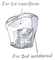
Gray's Anatomy (1918) plate, public domain
The second (intermediate) cuneiform is the smallest of the three cuneiforms, and its position is the clever part: it sits recessed between its neighbours, so the base of the second metatarsal locks into a mortise between the first and third cuneiforms. This recessed keystone is what makes the midfoot's Lisfranc joint so stable.
Key facts
- It is the smallest cuneiform.
- Its recessed position creates the mortise for the second metatarsal.
- This arrangement is the keystone of the transverse arch of the foot.
- The Lisfranc ligament ties it to the second metatarsal base.
- It rarely moves — and that stability is its function.
What can go wrong
The Lisfranc ligament anchoring this bone is the critical stabiliser of the midfoot; when it ruptures in sport or a fall, the second metatarsal base shifts and the arch is at risk. Such injuries are the reason persistent midfoot pain and bruising after a twist are X-rayed standing, under load.
Related procedures & tests
Related symptoms
Frequently asked questions
Why is the second cuneiform set back from the others?
So the second metatarsal can sit in a socket rather than on a flat line — like the keystone of an arch, it makes the whole midfoot resist being flattened under body weight.
What is the Lisfranc ligament?
A strong ligament from the second cuneiform to the base of the second metatarsal — the single most important soft-tissue lock of the midfoot joint complex.
Educational information only. This atlas explains normal anatomy and describes conditions in general terms; it is not medical advice and does not replace assessment by a qualified clinician. If you have symptoms that worry you, seek medical care.